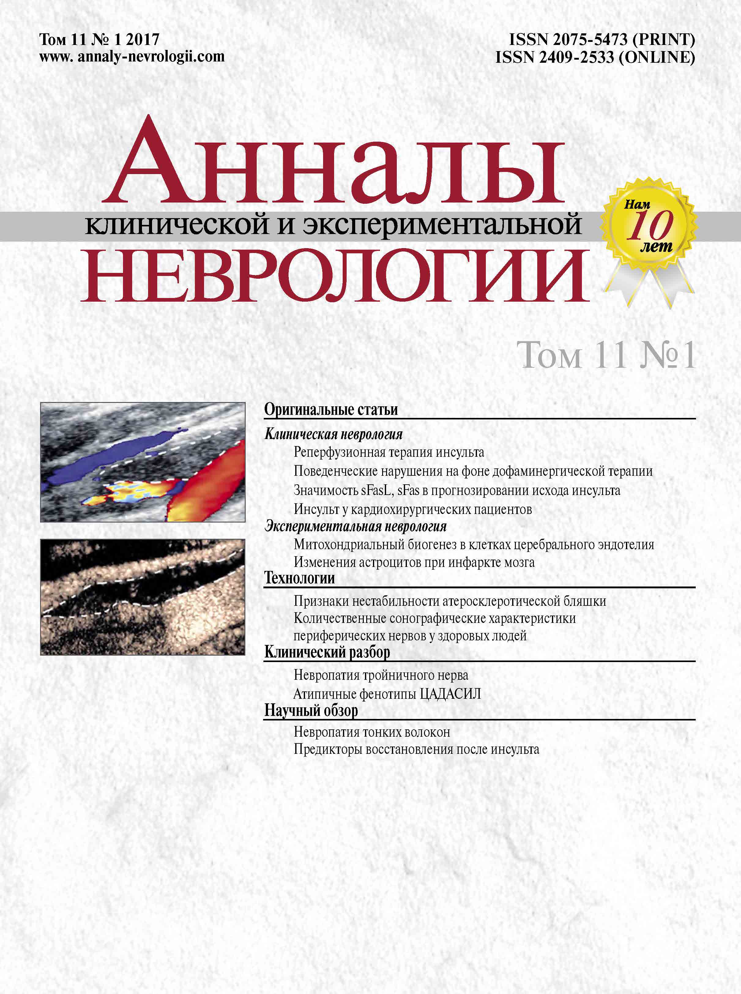
Том 11, № 1 (2017)
- Год: 2017
- Дата публикации: 12.05.2017
- Статей: 12
- URL: https://annaly-nevrologii.com/journal/pathID/issue/view/47
Весь выпуск
Оригинальные статьи
Персонификация подходов к реперфузионной терапии ишемического инсульта
Аннотация
Введение. Системный тромболизис рекомбинантным тканевым активатором плазминогена (rtPA) является «золотым стандартом» реперфузионной терапии у тщательно отобранных пациентов с ишемическим инсультом в первые 4,5 ч c момента развития симптомов заболевания.
Цель исследования – оценка клинических (выраженность неврологической симптоматики при поступлении) и лабораторных (показатели общего анализа крови) факторов, влияющих на прогноз заболевания после проведения системного тромболизиса.
Материалы и методы. Проспективно наблюдались 70 пациентов (48 мужчин и 22 женщины) в возрасте 61 [54; 69] лет с ишемическим инсультом, которым был проведен системный тромболизис rtPA в дозе 0,9 мг/кг. Забор крови для общего анализа (с подсчетом нейтрофилов и лимфоцитов) проводился до выполнения тромболитической терапии. Выраженность неврологических нарушений оценивалась с помощью шкалы инсульта Национальных институтов здоровья (NIH). Функциональный прогноз оценивали через 3 мес после инсульта по модифицированной шкале Рэнкина (mRS). Для выявления маркеров неблагоприятного прогноза острого периода инсульта (оценка по шкале mRS 3 балла и более) проведен ROC-анализ с определением чувствительности и специфичности.
Результаты. Выраженность неврологической симптоматики по шкале NIH при поступлении у пациентов составила 15 [11; 17] баллов. Время с момента развития неврологической симптоматики до поступления пациентов в стационар составило 138 [117; 170] мин, с момента поступления до начала системного тромболизиса («от двери до иглы») – 40 [30; 55] мин.
На основании ROC-анализа при оценке по шкале NIH при поступлении 12 баллов и более (чувствительность – 94%, специфичность – 57%), количестве нейтрофилов более 7,8•109/л (чувствительность – 45,5%, специфичность – 90,6%), лимфоцитов – менее 1,8•109/л (чувствительность – 81,8%, специфичность – 59,4%) можно прогнозировать неблагоприятный функциональный исход после системного тромболизиса.
Заключение. Персонифицированный подход к проведению системного тромболизиса может помочь прогнозировать его эффективность и способствовать разработке адекватных подходов к ведению пациентов. Пациенты с потенциально неблагоприятным прогнозом при системном тромболизисе могут быть целевой группой для выполнения механических методов реперфузии (тромбэкстракции).
 7-13
7-13


Поведенческие нарушения при болезни Паркинсона на фоне дофаминергической терапии
Аннотация
Введение. Болезнь Паркинсона (БП) – прогрессирующее неврологическое заболевание, в основе которого лежит дегенерация дофаминергических нейронов в компактной части черной субстанции. Наиболее эффективными в лечении этого заболевания являются дофаминергические препараты, которые могут приводить к развитию поведенческих расстройств или нарушений импульсного контроля (НИК). К НИК относят компульсивный шопинг, игроманию, гиперсексуальность, компульсивное переедание, пандинг (бесцельные повторяющиеся действия), дофаминовый дизрегуляционный синдром (ДДС).
Цель работы – выявление частоты поведенческих расстройств при БП с оценкой их влияния на показатели качества жизни и повседневной активности пациентов и их родственников.
Материалы и методы. Для определения распространенности НИК 340 пациентов с БП были подвергнуты анкетированию с помощью краткого опросника для выявления нарушений импульсного контроля – QUIP-Short. В последующий анализ были включены 60 больных БП с выявленными НИК (17% от общего числа обследованных) и 20 больных БП без поведенческих нарушений, у которых для оценки НИК, импульсивности, повседневной активности, качества жизни, тревоги, депрессии, когнитивных нарушений применялась батарея специальных тестов.
Результаты. ДДС имел место у 8% пациентов БП, пандинг – у 10%, компульсивное переедание – у 6%, гиперсексуальность – у 5%, компульсивный шопинг – у 4%, а игромания только у 1% пациентов. Показатель повседневной активности больных БП с НИК в среднем составил 60,05±9,76%, показатель качества жизни – 67,21±18,54%, причем эти значения были существенно ниже, чем у больных БП без поведенческих нарушений.
Заключение. НИК встречаются у каждого пятого больного БП на фоне дофаминергической терапии. Наиболее часто диагностируются ДДС и пандинг. Развитие НИК значительно влияет на повседневную активность больных и снижает качество жизни как больных, так и их родственников.
 14-20
14-20


Прогнозирование исхода острого периода ишемического инсульта: роль маркеров апоптоза
Аннотация
Введение. Апоптоз нервных клеток является не только следствием инфаркта головного мозга, он также представляет собой важное патогенетическое звено ишемического повреждения. Тем не менее роль апоптотических маркеров в прогнозировании функционального исхода острого периода ишемического инсульта (ИИ) не была установлена.
Цель работы – определение возможности прогноза исхода острого периода ИИ в максимально ранние сроки после его развития путем определения концентрации sFasL и sFas в периферической крови пациентов.
Материалы и методы. В условиях стационара обследованы 155 чел. Из них сформированы 3 группы: группа контроля – здоровые добровольцы (n=28)и две группы пациентов в зависимости от исхода острого периода ИИ – с благоприятным (балл по шкале оценки тяжести неврологического дефицита Национального института здоровья, NIHSS, на 21 сут менее 5) и с неблагоприятным исходом (балл по шкале NIHSS на 21 сут более 5). Концентрацию sFas, sFasL определяли на 1, 7 и 21 сут после ИИ методом ИФА, также вычисляли отношение sFasL к sFas.
Результаты. Показано, что если отношение концентраций sFasL и sFas равно или меньше 2,11±0,58, то прогноз благоприятный (NIHSS на 21 сут ≤5),если больше 2,11±0,58, то прогноз неблагоприятный (NIHSS на 21 сут >5).
Заключение. Предложенный способ имеет прогностическую значимость и высокую точность и может быть использован в клинической практике при определении стратегии ведения пациентов с ИИ.
 21-27
21-27


Внутрибольничный инсульт у пациентов кардиохирургического профиля
Аннотация
Введение. В структуре внутрибольничных инсультов многопрофильного стационара лидирующее положение занимают инсульты у пациентов кардиохирургического профиля. По данным литературы, частота инсульта от 0,2–0,4% для перкутанных операций на сердце до 16% после операций на клапанах сердца.
Цель исследования. Выявить факторы риска инсульта у пациентов кардиохирургического профиля, в т.ч. в зависимости от варианта оперативного вмешательства.
Материалы и методы. Группа исследования составила 58 случаев ОНМК у пациентов кардиохирургического отделения – 30,5% от общего количества зарегистрированных за 5-летний период (2011–2016 гг.) внутрибольничных инсультов.
Результаты. В группе исследования преобладал ишемический инсульт (54 пациентов; 93,1%, р<0,001), у четырех пациентов (6,9%) наблюдалась ТИА. Наибольшее число инсультов произошло у пациентов, перенесших шунтирующие операции на сердце (23 пациента, 41,1%) и операции по протезированию клапанов сердца – 25 случаев (44,6%), в 21,4% (12 случаев) ОНМК произошло после протезирования митрального клапана в сочетании с аннулопликацией трикуспидального клапана. Большинство инсультов развивалось в первые трое суток после оперативных вмешательств (36 пациентов, 64,3%, р<0,05).
Заключение. Пациенты кардиохирургического профиля, особенно после шунтирующих операций на сердце и операций по протезированию клапанов сердца, в первые трое суток требуют динамического контроля гемодинамики и тромбоэластограммы с целью профилактики инсульта. Несмотря на раннее выявление внутрибольничных инсультов, всем пациентам системный тромболизис был противопоказан. Для данной категории больных лечебной процедурой выбора должна рассматриваться механическая тромбэктомия.
 28-33
28-33


Активация лактатных рецепторов GPR81 стимулирует митохондриальный биогенез в клетках эндотелия церебральных микрососудов
Аннотация
Введение. Клетки церебрального эндотелия экспрессируют монокарбоксилатные транспортеры MCT1 для переноса лактата через гематоэнцефалический барьер (ГЭБ), регулируемые активностью CD147, а также рецепторы лактата GPR81 (HCAR1). Метаболизм и межклеточный транспорт лактата – важный механизм регуляции функциональной активности клеток ГЭБ.
Цель исследования. Изучить влияние активности GPR81 рецепторов в клетках церебрального эндотелия на экспрессию MCT1, CD147 и митохондриальную динамику, что позволит объяснить эффект локальной продукции лактата периваскулярными астроцитами на процессы ангиогенеза в ткани головного мозга.
Материалы и методы. В работе использовалась культура клеток церебральных эндотелиоцитов, выделенных из головного мозга 15–17 дневных эмбрионов крыс линии Wistar. Изучение митохондриального биогенеза церебральных эндотелиоцитов проводили по стандартному протоколу «MitoBiogenesis In-Cell ELISA Kit» (Abcam). Химическую гипоксию создавали путем инкубации в присутствии 50 мкМ йодацетата в течение 30 мин. В качестве агониста рецепторов лактата GPR81 использовали 3Cl-5OH-BA (Calbiochem) в концентрации 5, 50 и 500 мкМ в течение 24 час. Количество клеток, экспрессирующих молекулы GPR81, CD147 и MCT1, оценивали с использованием двойного непрямого метода иммуноферментного окрашивания.
Результаты. Впервые обнаружено, что длительная стимуляция GPR81 рецепторов 3Cl-5OH-BA в дозозависимой манере приводит к интенсификации митохондриального биогенеза (до 1,5 раз, р<0,05). В то же время зафиксировано статистически значимое (р<0,05) подавление экспрессии монокарбоксилатных транспортеров MCT1 в опытной группе по сравнению с контрольной (с 81±1,6% до 40,7±4,4%) и сопряженного с ними белка CD147 (с 57,4±3,3% до 48,3±2,9%) в церебральных эндотелиоцитах.
Заключение. Полученные данные расширяют спектр возможных приложений действия агонистов GPR81 для модуляции межклеточных взаимодействий в нейроваскулярной единице и контроля функциональной активности клеток эндотелия церебральных микрососудов.
 34-39
34-39


Иммуноцитохимические и морфометрические изменения астроглии в перифокальной зоне моделируемого инфаркта мозга
Аннотация
Введение. Перифокальная зона (ПЗ) инфаркта головного мозга содержит гибнущие и реактивно измененные нейроны, судьба которых зависит от характера межклеточных взаимодействий и, в частности, от реакции астроцитов, участвующих как в повреждении нейронов, так и в нейропротекции. Особенности реакции астроцитов на ишемическое повреждение и значение их активации при глиозе изучены недостаточно.
Цель исследования. Методами иммуноморфологии и компьютерной морфометрии оценить изменения астроглии в перифокальной зоне инфаркта мозга в зависимости от сроков его воспроизведения.
Материалы и методы. Инфаркт моделировали в левом полушарии коры головного мозга крыс (n=10) окклюзией средней мозговой артерии. Оценивали распределение и форму астроцитов на 3-й и 21-й дни после операции, исследовали локализацию кислого глиофибриллярного белка (GFAP), аквапорина-4 (AQP4) и глутаминсинтетазы (GlnS) в перифокальной зоне.
Результаты. Параметры формы, распределение астроцитов и экспрессия GFAP значимо менялись в зависимости от срока и расстояния до очага повреждения. На 3-й день площадь, занимаемая отростками астроцитов, снижалась на 15% от контроля, а на 21-й день – возрастала на 35%. Экспрессия GlnS и AQP4 на 3-й день вблизи очага инфаркта снижалась, а на 21-й день наблюдали противоположные изменения. Также выявили перераспределение исследованных белков в отростках реактивных астроцитов. Выделили два морфологических типа астроцитов: рубец-формирующие поляризованные астроциты, характеризующиеся перераспределением маркерных белков в отростках, и транзиторно-активированные, отличающиеся умеренными изменениями.
Заключение. Выявлена гетерогенность астроцитов в ПЗ инфаркта и зависимость их структурно-функциональных изменений от расстояния до очага повреждения и сроков после инфаркта. При помощи иммуногистохимического и морфометрического анализа охарактеризованы рубец-формирующие и транзиторно-активированные астроицты, имеющие разное значение для ремоделирования и репарации ишемизированной нервной ткани в перифокальной зоне инфаркта.
 40-46
40-46


Обзоры
Невропатия тонких волокон
Аннотация
Невропатия тонких волокон (НТВ), несмотря на тридцатилетнюю историю изучения, остается одним из самых загадочных заболеваний, которые с трудом поддаются диагностике и лечению. Распространенность НТВ составляет 52,95 на 100 000 населения, наиболее частой причиной ее развития считается сахарный диабет. В результате повреждения тонких миелинизированных Аδ- и немиелинизированных С-волокон формируются хронический невропатический болевой синдром, расстройства температурной чувствительности, вегетативные нарушения. Заболевание в основном развивается по «восходящему типу», распространяясь со стоп на проксимальные отделы и руки, тип поражения нервов – первично аксональный. Несмотря на то, что НТВ считается одной из самых «доброкачественных» невропатий, т.к. не вовлекает крупные чувствительные и двигательные волокна, качество жизни пациентов заметно снижается.
 73-79
73-79


Прогностические факторы восстановления нарушенных в результате ишемического инсульта двигательных функций
Аннотация
Проблема поиска прогностических факторов восстановления нарушенных функций после инсульта имеет большую актуальность в связи с высокой распространенностью острых нарушений мозгового кровообращения (ОНМК). Тяжелые инвалидизирующие последствия инсульта связаны с огромными экономическими потерями во всем мире, что усугубляется отсутствием индивидуального подхода к восстановлению с учетом клинических данных и нейропластических особенностей мозга каждого пациента. Несмотря на внедрение современных диагностических методов, в том числе нейровизуализационных, многие потенциальные предикторы восстановления утраченных функций после церебральной катастрофы остаются невыясненными или неуточненными. В статье приведен обзор наиболее изученных прогностических факторов восстановления утраченных функций после ишемического инсульта.
 80-89
80-89


Технологии
Новые подходы к оценке признаков нестабильности атеросклеротической бляшки в сонных артериях
Аннотация
Введение. Использование контрастных препаратов при ультразвуковом исследовании сосудов стало новым направлением в неинвазивной оценке признаков нестабильности атеросклеротической бляшки (АСБ), к важнейшим из которых относится характер ее неоваскуляризации. Однако остаются нерешенными вопросы, касающиеся точности методов количественной оценки неоваскуляризации бляшки.
Цель исследования. Оценить признаки нестабильности АСБ в сонных артериях по данным дуплексного сканирования с контрастным усилением с разработкой собственного подхода к количественной оценке неоваскуляризации.
Материалы и методы. В исследование вошли 26 больных с каротидным атеросклерозом, которым была выполнена каротидная эндартерэктомия (n=27) с последующей морфологической верификацией бляшек. Всем пациентам выполнялось стандартное дуплексное сканирование и сканирование с введением контрастного вещества «Соновью».
Результаты. Неоваскуляризация выявлена во всех 27 АСБ по данным патоморфологического и ультразвукового исследования с контрастированием. Общее количество сосудов на 1 см2 бляшки по результатам ультразвукового исследования составило 6–51 [21±14/см2], по результатам патоморфологического исследования – 19–1224 [236±249/см2]. Согласно результатам ультразвукового исследования, абсолютные значения находились вблизи величины плотности сосудов диаметром ≥30 мкм в бляшке, определенной при патоморфологическом исследовании, и значимо от нее не отличались (р=0,67). Сосуды диаметром <20 мкм, составлявшие до 96% от всех микрососудов АСБ по морфологическим данным, при ультразвуковом исследовании не определяются. В одном случае изъязвление поверхности АСБ удалось выявить только после введения контрастного вещества. Наибольшие сложности при ультразвуковой оценке неоваскуляризации представляли бляшки с включением кальция различной степени выраженности.
Заключение. Ультразвуковое исследование с контрастированием может быть использовано как информативный метод неинвазивного определения признаков нестабильности АСБ, позволяющий достаточно точно оценивать неоваскуляризацию при диаметре микрососудов ≥30 мкм. Наличие кальция в АСБ может значительно влиять на результаты исследований.
 47-54
47-54


Количественные сонографические характеристики периферических нервов у здоровых людей
Аннотация
Введение. Ультразвуковое исследование (УЗИ) позволяет неинвазивно сканировать периферические нервы с получением количественных и качественных характеристик.
Цель исследования. Определить нормативные значения площади поперечного сечения (ППС) нервов рук и ног, а также спинномозговых нервов у здоровых добровольцев.
Материалы и методы. Проведено УЗИ здоровым добровольцам – 40 мужчинам и 40 женщинам, средний возраст 40,3±15,1 (от 18 до 70 лет) – периферических нервов рук и ног, а также спинномозговых нервов плечевого сплетения с обеих сторон. Использовался ультразвуковой сканер «Sonoscape S20» (Китай) с линейным датчиком 8–15 МГц. Оценивалась ППС периферических нервов. В анализ взяты рост, вес, индекс массы тела (ИМТ), возраст, пол.
Результаты. Получены нормативные значения ППС основных нервов рук, ног и спинномозговых нервов плечевого сплетения. Не обнаружено достоверной корреляции основных антропометрических параметров (рост, вес, ИМТ), а также возраста и пола с величиной ППС периферических нервов и плечевого сплетения.
Заключение. Получены данные, сопоставимые с результатами измерений других авторов, что свидетельствует об общности методических подходов к УЗИ нервов, принятых в других лабораториях.
 55-61
55-61


Клинический разбор
Болезненная невропатия тройничного нерва, обусловленная опоясывающим герпесом
Аннотация
Наиболее частым проявлением опоясывающего герпеса является невропатия глазного нерва (I ветви тройничного нерва). Невропатия глазного нерва встречается в 20% случаев опоясывающего герпеса. При невропатии тройничного нерва выделяют три типа боли: постоянную жгучую боль, приступообразную боль, боль, возникающую при неболевом раздражении. На коже выявляются области гипестезии, анестезии, дизестезии. Постгерпетическая невропатия характеризуется болью длительностью 3 мес и более от момента появления герпетических высыпаний. Комбинированная терапия, включающая раннее применение противовирусных средств и трициклических антидепрессантов, является наиболее эффективной.
 62-67
62-67


Атипичные клинические случаи церебральной аутосомно- доминантной артериопатии с субкортикальными инфарктами и лейкоэнцефалопатией (ЦАДАСИЛ)
Аннотация
Церебральная аутосомно-доминантная артериопатия с субкортикальными инфарктами и лейкоэнцефалопатией (ЦАДАСИЛ) – наследственное заболевание центральной нервной системы c передачей по аутосомно-доминантному типу, обусловленное мутациями гена NOTCH3. В классических случаях ЦАДАСИЛ проявляется головными болями, повторными нарушениями мозгового кровообращения и прогрессирующим когнитивным снижением. Важное диагностическое значение имеет церебральная магнитно-резонансная томография, выявляющая множественные лакунарные инфаркты в области базальных ядер, ствола мозга и мозжечка, очаговые изменения белого вещества и диффузные изменения по типу лейкоареоза. Изредка ЦАДАСИЛ может проявляться иными симптомами и протекать под маской необычных для данного заболевания фенотипов. Мы приводим два генетически подтвержденных случая ЦАДАСИЛ с атипичной клинической картиной, манифестировавших в виде тремора преимущественно мозжечкового либо эссенциального типа в сочетании с когнитивными и аффективными нарушениями. Обсуждаются основные принципы диагностики данногоклинически полиморфного заболевания.
 68-72
68-72


