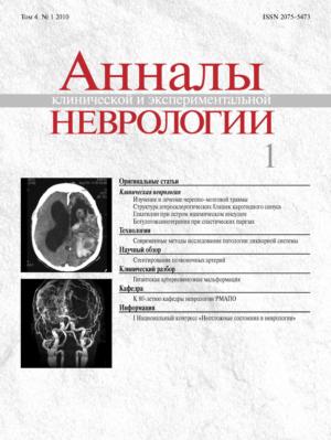
Vol 4, No 1 (2010)
- Year: 2010
- Published: 13.03.2010
- Articles: 7
- URL: https://annaly-nevrologii.com/journal/pathID/issue/view/34
Full Issue
Original articles
Modern approaches to the management of traumatic brain injury
Abstract
The authors report on the problem of Traumatic Brain Injury (TBI) and main ways of its solution. Special emphasis is being placed on literature data bank and personal experience in studying and making precise diagnosis of diffuse and focal brain damage using Diffuse-tensor MRI (DT-MRI) and its other modalities. A comparative analysis of DT-MRI findings in 8 healthy volunteers and 22 patients in coma with severe diffuse axonal injury (DAI) at the period of 2–17 days after trauma demonstrated significant changes in the corpus callosum and corticospinal tracts (CST) caused by DAI. Fractional anisotropy was considered the most sensitive indicator of their damage in the early stage of DAI. The authors developed and described a new approach to the management of brain concussion which allows out-patient treatment of such patients provided there is no focal damage at GCS 15 and on condition thorough neurological and regular CT- and MRI examinations are performed.
 4-12
4-12


Structure of carotid sinus atherosclerotic plaques and disturbances of cerebral blood circulation
Abstract
A morphological study of 200 atherosclerotic carotid sinus (CS) plaques obtained at carotid endarterectomy revealed the structural components and processes characteristic for severe atherosclerosis: foci of atheromatosis and edema, necrosis of collagen and elastic fibres, newly formed vessels and hemorrhages of various duration, lipophages and lymphocytes, portions of fibrosis and calcification, covering thinning and ulceration, thrombi and the contents of plaques (atheromatous masses, cholesterol crystals, lipophages, calcificates) on their surface. Clinical and morphological comparisons indicated that patients with ischemic stroke in their history had more severe atherosclerosis than those with asymptomatic CS stenosis. They were more commonly found to have vulnerable atherosclerotic plaques that showed a predominance of atheromatosis foci over the portions of fibrosis and calcification, covering thinning and destruction, formation of thrombi on its surface. It was ascertained that concomitant arterial hypertension might play a crucial role in the pathogenesis of hemorrhages into the atherosclerotic CS plaque.
 13-19
13-19


Multicentral pilot clinical trial of gliatilin in treatment of acute ischemic stroke
Abstract
The multicentral pilot clinical trial of gliatilin in treatment of acute ischemic stroke was held in 2006–2008 in Russia. The trial involved 122 patients, who received basic treatment and gliatilin during 3 months after the stroke onset (1–15 days – 2000 mg per day, 16–30 days – 1000 mg per day, 31–90 days – 800 mg per day). All patients underwent clinical examination (including repeated NIHSS, Rankin scale, Barthel index assessment), laboratory and ultrasound examination, computer tomography or magnetic resonance imaging during 24 hours after the stroke onset. 25 patients underwent repeated multimodal magnetic resonance imaging (T1-, T2-, diffusion-, perfusion-weighted imaging). The results of examination of patients who received basic treatment alone were taken from literature. The trial results show, that treatment of acute ischemic stroke with gliatilin promotes neurological recovery and increase of patient’s self-care ability, which is probably concerned with the smaller final volume of brain infarct.
 20-28
20-28


Application efficiency and effectiveness of Botulinum neurotoxin type A in complex treatment of patients with post – stroke spasticity in arm
Abstract
Studying of efficiency of application botulinum neurotoxin type A in complex treatment 51 patients with poststroke spastic hemiparesis with mean time from stroke onset 33.8 months. It was observed severe spastisity (mean of scale of Ashworth – 3.3+1.0) at all patients in the neurologic status. Patients were divided in three groups. The first group (n = 14) received botulinum neurotoxin type A in a complex with standard restoring therapy (BtxA+SRT). In the second group (n = 18) the patients received BtxA+SRT in a complex with specialized functional training using device «ARMEO». The third group (n = 19) received only BtxA. Comparisons of scores on the MAS, AS, FIM and goniometry at baseline, week 5 and the 8-month follow-up are reviled the most considerable improvement of hand function in second group which received BtxA+SRT in a complex with specialized functional training using device «ARMEO». We believe that BtxA must use with complex therapy including the functional training of hand with device «ARMEO».
 29-33
29-33


Reviews
Vertebral artery stenting: problem of restenosis
Abstract
Balloon angioplasty with stenting is actual uninvasive and safe method of vertebral arteries stenosis treatment, having high level of technical success. But high rate of restenosis development is leading problem of endovascular intervention. In review the problem of restenosis development after vertebral artery proximal part stenting is being discussed as well as its clinical onset, possible cause and factors promoting its formation and probable direction of this problem solving.
 41-48
41-48


Technologies
Modern methods of investigation CSF pathology
Abstract
Modern MRI (Magnetic Resonance Imaging) programme support modification more and more often makes a radiologist to refuse the invasive techniques in favour of more safe methods like Magnetic Resonance Myelography, Magnetic Resonance Cisternography, combination of CT and MR-cisternography. To evaluate CSF flow and obtain quantitative characteristics of linear and regional CSF flow the method of phase-contrast MRI is used. Today these methods have become a routine practice and may be indicated for all patients with the corresponding CSF system pathology. The spectrum of diagnoses is rather large: all types of hydrocephalus, arachnoid cysts, midline tumours and tumors located in the CSF lumen, «empty» saddle, different types of CSF leakage, Arnold-Chiari malformations, anomalous development of the brain and brain ventricles, ventriculostoma (artificial and spontaneous), postoperative CSF collections convexital hygromas. Magnetic Resonance Myelography and Magnetic Resonance Cisternography can successfully replace the invasive methods of visualization of cerebral and spinal CSF spaces. Phase-contrast MRI has proved to be efficient in demonstrating open hydrocephalus and providing postoperative control in Arnold-Chiari malformations.
 34-40
34-40


Clinical analysis
Congenital huge arterio-venous malformation
Abstract
Arterio-venous malformation represent congenital anomaly of vessels at which inside a brain tissue texture of pathological arteries and veins are formed. Under the world data frequency of occurrence АВМ makes from 0.89 till 1.24 on 100 000 population in a year. The lethality is 10–15%, death reason is usually the development of hemorrhage. Rupture of arteriovenous malformation usually occurs at the age of 20–40 years and often is the first display of disease. In rare instances arterio-venous malformation happens so big that causes an ischemia of the next sites of a brain and clinically proceeds like a brain tumour. More often such malformations meet in pool of an average brain artery and extend from a bark of a brain to ventricules, often accompanied by a hydrocephaly. The clinical case of a favorable outcome of repeated subarachnoid hemorrhages at the patient with congenital huge arteriovenous malformation under conservative therapy is described.
 49-52
49-52


