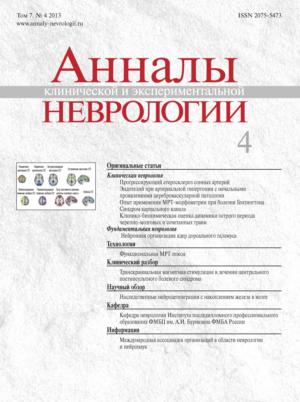
Том 7, № 4 (2013)
- Год: 2013
- Дата публикации: 09.12.2013
- Статей: 9
- URL: https://annaly-nevrologii.com/journal/pathID/issue/view/16
Весь выпуск
Оригинальные статьи
Прогрессирующий церебральный атеросклероз: клинические, биохимические и морфологические аспекты
Аннотация
В статье обсуждаются основные патогенетические механизмы прогрессирования атеросклеротического поражения магистральных артерий головы, приводящие к реализации ишемических нарушений мозгового кровообращения. Рассматриваются ультразвуковые, морфологические и биохимические предикторы изменений структуры атеросклеротической бляшки (АСБ). Многоплановость патогенетических моментов развития и прогрессирования церебрального атеросклероза как ведущей причины развития ишемических поражений мозга диктует необходимость изучения различных аспектов этой проблемы – от структурно-морфологических изменений сосудов и вещества мозга до метаболических параметров и различных характеристик крови (биохимических, липидологических, конформационных, гемостатических).
 4-9
4-9


Функция эндотелия у больных с начальными клиническими проявлениями хронической цереброваскулярной патологии при артериальной гипертонии
Аннотация
Профилактика развития и прогрессирования хронической цереброваскулярной патологии (ХЦВП) является приоритетным направлением предупреждения цереброваскулярных заболеваний (ЦВЗ). При этом особое внимание уделяется начальным клиническим проявлениям данной патологии, в развитии которой особое место занимает дисфункция эндотелия. Целью исследования явился анализ особенностей состояния функции эндотелия у пациентов с началь ными проявлениями ХЦВП при неосложненной артериальной гипертонии (АГ). Комплексное унифицированное обследование прошли 48 мужчин и 61 женщина (средний возраст 57,4±5,8 лет). Исключались пациенты, перенесшие острое нарушение мозгового кровообращения (ОНМК) или острый коронарный синдром (ОКС), черепно-мозговую травму или тяжелое соматическое заболевание, а также имеющие стеноз магистральных артерий головы (МАГ) более 30%. Обследование включало наряду с клиническим и неврологическим осмотром проведение УЗИ,Эхо-КГ, СМАД, МРТ/КТ, комплексное нейропсихологическое обследование. Функция эндотелия исследовалась с помощью функциональных проб (гемореологическая и вазоактивная манжеточная проба (МП), ЦВР с нитроглицерином). В качестве биохимических маркеров состояния функции эндотелия использовались фактор фон Виллебранда (ФВ) и гомоцистеин (ГЦ). Получены следующие данные: установлена значительная частота нарушения функции эндотелия у лиц с неосложненной АГ (64%), показана высокая частота совпадения результатов обеих МП. Выявлена зависимость результатов гемореологической МП от исходных показателей. Для оценки прогностического значения дисфункции эндотелия в отношении прогрессирования ХЦВП целесообразна организация проспективного наблюдения за когортой.
 10-15
10-15


Опыт применения МРТ-морфометрии при болезни Гентингтона
Аннотация
Для одного из наиболее значимых наследственных нейродегенеративных заболеваний – болезни Гентингтона (БГ) – характерна церебральная атрофия, характер которой нуждается в уточнении. Технология МРТ-морфометрии позволяет количественно оценить атрофию различных областей головного мозга, что позволяет рассматривать его как потенциальный биомаркер нейродегенерации. Мы применили у 24 пациентов с БГ, 10 доклинических носителей мутации и 9 здоровых лиц две разновидности метода МРТ-морфометрии – сравнение мозговых объемов в целом и подсчет объемов заранее заданных областей интереса. При сравнении мозговых объемов в целом у пациентов с БГ хвостатое ядро и скорлупа с двух сторон, преи постцентральная извилины были достоверно меньше, чем у лиц из группы контроля, в то время как при сравнении заданных регионов интереса уменьшение объема у больных затронуло хвостатое ядро, бледный шар и скорлупу с двух сторон. У клинически здоровых носителей мутантного гена бледный шар, скорлупа, пре- и постцентральная извилины билатерально оказались достоверно больше, чем у пациентов, а бледный шар слева – меньше, чем в контроле. Выявлены большее поражение доминантной стороны у пациентов и у носителей гена БГ, а также отрицательные корреляции объемов подкорковых и корковых структур с тяжестью мутации, двигательными и когнитивными нарушениями.
 16-19
16-19


Особенности диагностики синдрома карпального канала с помощью электромиографии и ультразвукового исследования
Аннотация
Синдром карпального канала (СКК) представляет собой заболевание, характеризующееся чувствительными и двигательными проявлениями, связанными с компрессией срединного нерва (СН) на уровне запястья. Целью настоящего исследования явилось сравнение двух диагностических методов – электромиографии (ЭМГ) и ультразвукового исследования (УЗИ) – для установления наиболее точного и надежного способа диагностики СКК. Были исследованы 67 пациентов с клиническим диагнозом СКК и 42 здоровых добровольца в качестве группы контроля. Для методов ЭМГ и УЗИ были вычислены показатели чувствительности и специфичности, а также определена степень поражения СН с помощью этих технологий. Чувствительность и специфичность для ЭМГ составила 93% и 97%, а для УЗИ – 85% и 73% соответственно. Степени поражения СН, определенные с помощью ЭМГ и УЗИ, не соответствовали друг другу. По нашим данным, метод ЭМГ обладает большей чувствительностью в сравнении с УЗИ при диагностике СКК. Метод ЭМГ также позволяет количественно оценивать степень поражения СН, в то время как УЗИ дает преимущественно качественную оценку поражения СН при СКК.
 20-26
20-26


Клинико-биохимическая оценка динамики острого периода черепно-мозговых и сочетанных травм
Аннотация
Целью работы являлось изучение клинических эффектов и динамики изменения параметров системы перекисного окисления липидов (ПОЛ) и антиоксидантной защиты при лечении пациентов в остром периоде черепно-мозговых и сочетанных травм пептидным препаратом кортексин. В результате работы выявлена способность препарата достоверно снижать интенсивность процессов ПОЛ при черепно-мозговой травме. Клинический эффект выражался в уменьшении астенических проявлений, заметном расширении объема внимания и памяти, повышении работоспособности, увеличении темпа психической деятельности, что подтверждалось данными психометрических тестов. Установлено, что применение кортексина в остром периоде черепно-мозговой и сочетанной черепно-лицевой травмы значительно повышает качество жизни пациентов, оказывает седативный и антидепрессивный эффект, снижает эпилептическую активность и уменьшает частоту эпилептических приступов.
 27-31
27-31


Транскраниальная магнитная стимуляция в лечении центрального постинсультного болевого синдрома
Аннотация
Транскраниальная магнитная стимуляция (ТМС) – метод, основанный на возбуждении нейронов головного мозга переменным магнитным полем.
В последние годы появились сообщения об эффективности высокочастотной ТМС при лечении фармакорезистентного центрального постинсультного болевого синдрома (ЦПБС). Представленный клинический разбор описывает двух пациентов с клиникой ЦПБС с разной локализацией и объемом очага поражения. ТМС применялась в режиме высокочастотной стимуляции (10 Гц) на первичную моторную кору пораженного полушария. После стимуляции зафиксировано значимое снижение выраженности болевого синдрома по визуальной аналоговой шкале (ВАШ). Показано, что после окончания ТМС боль постепенно возвращается к прежнему уровню через 3-4 месяца.
 45-50
45-50


Обзоры
Общие принципы нейронной организации ядер дорсального таламуса человека
Аннотация
В ядрах дорсального таламуса (VA, VL, AV, AM, AD, MD) человека изучены нейроны методом Гольджи. Метод позволил идентифицировать виды длинноаксонных густоветвистых нейронов (I тип): кустовидные и кустовидные шипиковые среднего размера, крупные и гигантские кустовидные, древовидные, кисточковые, двухпучковые и переходные формы нейронов. Идентифицированы также виды длинноаксонных редковетвистых: короткодендритные и ретикулярные нейроны. II тип нейронов – короткоаксонные нейроны (интернейроны) – представлен гладкодендритными, «лохматодендритными» и длиннодендритными клетками. Дана классификация нейронов ядер дорсального таламуса, установлены закономерности внутренней организации этих ядер.
 32-38
32-38


Наследственные нейродегенерации с накоплением железа в мозге
Аннотация
Нейродегенерации с накоплением железа в мозге (ННЖМ) – клинически и генетически гетерогенная группа наследственных (преимущественно аутосомно-рецессивных) прогрессирующих болезней ЦНС с общим признаком – накоплением железа в базальных ганглиях, дающим характерную картину при нейровизуализации. В настоящее время идентифицировано 9 генов, связанных с разными ННЖМ, часть из этих генов обусловливают развитие несколько аллельных фенотипов. В обзоре суммированы современные клинические и молекулярно-генетические данные о ННЖМ, особенно о новых формах и атипичных клинических вариантах.
 51-60
51-60


Технологии
Функциональная магнитно- резонансная томография покоя: новые возможности изучения физиологии и патологии мозга
Аннотация
В последнее время с целью изучения основных сенсорных, эмоциональных и когнитивных процессов в норме и при патологии был предложен новый метод – функциональная магнитно-резонансная томография в состоянии покоя (фМРТп). Он позволяет оценить степень спонтанной коактивации различных центров ЦНС в покое на основе сходства временных характеристик нейрональной активности, выявляемой для анатомически удаленных друг от друга участков головного мозга. При фМРТп-исследованиях показано существование стабильных функционально связанных «сетей покоя» головного мозга, изучение которых перспективно в контексте анализа фундаментальных механизмов развития неврологических заболеваний. Нами впервые в России было проведено фМРТп-исследование в группе из 10 здоровых субъектов и выявлен отчетливый паттерн сети пассивного режима работы головного мозга, согласующийся по своему характеру с данными зарубежных исследований. Исследование с помощью фМРТп интегративной системы функционально взаимодействующих участков головного мозга человека может помочь по-новому взглянуть на широкие нейрональные взаимосвязи в рамках центральной нервной системы.
 39-44
39-44


