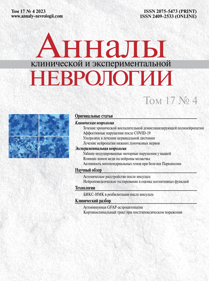
Vol 17, No 4 (2023)
- Year: 2023
- Published: 25.12.2023
- Articles: 12
- URL: https://annaly-nevrologii.com/journal/pathID/issue/view/80
Original articles
The Long-Term Course of Chronic Inflammatory Demyelinating Polyneuropathy: a Retrospective Study
Abstract
Introduction. Chronic inflammatory demyelinating polyneuropathy (CIDP) is characterized by long-term progressive or relapsing course, neurological deficit, and disability of varied severity. The course of CIDP after specific therapy and, if necessary, long-term maintenance treatment are to be studied.
Objective: To evaluate CIDP clinical and history characteristics over the long-term follow-up (> 5 years), to compare long-term CIDP course in a number of clinical variants and onset types, and to determine clinical predictors of unfavorable CIDP course.
Materials and methods. The study included 45 patients diagnosed with CIDP based on EAN/PNS 2021 criteria lasting for 5 or more years. Retrospective collection and analysis of medical records and clinical history were performed. Internationally accepted scales were used to assess neurological deficit (NIS, MRCss), disability (INCAT), and disease activity status (CDAS). The criteria of unfavorable course were developed to evaluate factors affecting CIDP course.
Results. Among the patients with CIDP history of >5 years, each third (34%) had no neurological deficit and remained in long-term clinical remission (CDAS 1). The vast majority (90%) responded to first-line therapy in early disease, while only 53% of patients required maintenance treatment in 5 or more years of the onset. With the developed criteria (poor response to glucocorticosteroids (GCS), need for maintenance therapy, and CDAS 3–5), unfavourable CIDP course was detected in 24 (53.3%) participants. Its probability increased in later onset (47 [30; 50] years), the chronic type of onset, and delayed specific therapy. The most significant predictors included low total NIS score at onset (<60 points) and multifocal CIDP.
Conclusions. The course of typical CIDP is relatively favorable if timely diagnosed, and pathogenetic treatment initiated. Patients with acute and subacute onset demonstrate the best long-term status. The predictors of unfavourable disease course include mild neurological deficit at onset (NIS total score <60 points) and multifocal CIDP.
 5-16
5-16


Mood Disorders After COVID-19
Abstract
Introduction. The COVID-19 pandemic has led to a high prevalence of post-COVID-19 syndrome (PCS), with mood disorders being the most common manifestations.
Objective: To study the prevalence of PCS-associated mood disorders and their features.
Materials and methods. We examined patients after COVID-19 (n = 91; age: 24-84 years; median time to recovery: 7 months) using the following tools: the BDI and HADS (screening for anxiety and depression); the Starkstein Apathy Scale; FIS and FSS (fatigue assessment); the MoCA, MMSE, and FAB (cognitive assessment); the FIRST, ESS, PSQI, and ISI (sleep disorders evaluation); the EQ5D (quality of life measurement). We designed a special questionnaire to collect data related to a history of COVID-19 and patients' condition after discharge. In addition, we analyzed electronic medical records and discharge summaries and performed neurological examination.
Results. Of all the examined patients, 65 (71.4%) participants had signs and symptoms of PCS. Mood disorders were observed in 33 (50.8%) cases, with apathy (78.7%), anxiety (66.7%), and fatigue (60.6%) being the most common. Depressive disorders were found in 12 (36.3%) patients. Cognitive functions were impaired in 7 (21.2%) patients; sleep disorders were observed in 16 (48.5%) cases. We found a positive correlation between depressive disorders and fatigue based on the BDI, FIS, and FSS scores (rS = 0.711; rS = 0.453), depressive disorders and anxiety (rS = 0.366), fatigue and apathy (rS = 0.350). Anxiety increased the risk of sleep disorders (rS = 0.683). Quality of life has been shown to decrease in patients with mood disorders due to the negative effect of long-term fatigue and depressive disorders.
Conclusions. There is a close connection between different types of mood disorders that develop after COVID-19 and exacerbate symptoms of each other. Early diagnosis and treatment of these disorders can improve patients' quality of life and preserve their ability to work.
 17-27
17-27


MRI-Guided Focused Ultrasound in Cervical Dystonia
Abstract
Introduction. MRI-guided focused ultrasound (MRgFUS) is approved for management of various movement disorders, primarily essential tremor and Parkinson’s disease (PD), with favorable long-term outcomes in numerous patients worldwide. However, few case studies describe the use of this modality for symptomatic treatment of dystonias that, as the third most common movement disorder, may be rather disabling.
Objective: To improve outcomes in patients with cervical dystonia (CD) using MRgFUS.
Materials and methods. We retrospectively analyzed 13 cases of various CD types managed with MRgFUS in single or multiple sessions. The mean age of the patients was 42 [39; 53] years. The Toronto Western Spasmodic Torticollis Rating Scale (TWSTRS) was used to assess patients' statuses and severity of CD symptoms during therapy and the last available observation period. The targets included the pallidothalamic tract and the thalamic ventral oralis complex nucleus or their combination.
Results. The mean follow-up period was 13.3 ± 3.4 months (July 2021 to April 2023). The mean CD severity sum score (TWSTRS score) was 22 [16; 25] before MRgFUS and 6 [4; 9] in the last observation. Therefore, we report 70.6% [55.6; 76.5] improvement (paired samples t-test p = 0.0025).
Conclusion. Available data evidence that MRgFUS is efficient and sufficiently safe for symptomatic treatment in pharmacoresistant CD patients. A number of vital aspects of MRgFUS have to be specified in larger CD cohorts in the long-term follow-up.
 28-34
28-34


Long-Term Outcomes of Management of Inferior Alveolar Neuropathy Following Orthognatic Surgeries in Patients with Mandibular Anomalies and Deformities
Abstract
Introduction. Orthognatic surgery is a routine method to manage mandibular anomalies and deformities.
Objective: To assess long-term outcomes of rhythmic peripheral magnetic stimulation (rPMS) in patients with neuropathy of the inferior alveolar nerve (IAN) resulting from the surgical treatment of mandibular anomalies and deformities.
Materials and methods. The study included 8 males and 16 females aged 32 ± 12 years with IAN neuropathy following the surgical treatment of mandibular anomalies and deformities. Therapeutic rPMS was performed with the Neuro-MS magnetic stimulator (Neurosoft, Ivanovo, Ivanovo Region, Russian Federation). Trigeminal and brainstem acoustic evoked potentials (EPs) were registered with Neuro-MVP (Neurosoft) to assess rPMS both at baseline (in 10 days) and in long term (in 18 ± 2 months).
Results. Sensory disorders and pain prevailed in postoperative IAN neuropathy. Sensory disorders improved in 20 patients following 10-day rPMS. The clinical effect persisted in re-assessment. In long term, acoustic brainstem EPs normalized and trigeminal EPs did not change negatively.
Conclusion. The use of rPMS in IAN neuropathy following orthognatic surgeries contributes to the functional improvement and stabilization of the peripheral and central brainstem and the trigeminal system.
 35-39
35-39


Long-term Intracerebroventricular Administration of Ouabain Causes Motor Impairments in C57Bl/6 Mice
Abstract
Introduction. Cardiac glycosides are natural ligands of Na+/K+-ATPase, which regulate its activity and signaling. Intracerebroventricular administration of ouabain has been previously shown to induce hyperlocomotion in C57Bl/6 mice via a decrease in the rate of dopamine reuptake from the synaptic cleft.
Materials and methods. This study involved forty C57BL/6 mice. 1.5 μL of 50 μM ouabain was administered daily into the left lateral cerebral ventricle over the course of 4 days. On day 5, open field, beam balance, and ladder rung walking tests were performed to assess the locomotor activity and motor impairments in the mice. We evaluated changes in the activation of signaling cascades, ratios of proapoptotic and antiapoptotic proteins, and the amount of α1 and α3 isoforms of the Na+/K+-ATPase α-subunit in brain tissue using Western blotting. Na+/K+-ATPase activity was evaluated in the crude synaptosomal fractions of the brain tissues.
Results. We observed hyperlocomotion and stereotypic behavior during the open field test 24 hours after the last injection of ouabain. On day 5, the completion time and the number of errors made in the beam balance and ladder rung walking tests increased in the mice that received ouabain. Akt kinase activity decreased in the striatum, whereas the ratio of proapoptotic and antiapoptotic proteins and the number of Na+/K+-ATPase α-subunits did not change. Na+/K+-ATPase activity increased in the striatum and decreased in the brainstem.
Conclusions. Long-term exposure to ouabain causes motor impairments mediated by changes in the activation of signaling cascades in dopaminergic neurons.
 40-51
40-51


Copper Ions Reduced Toxicity of Sodium Azide and Lipopolysaccharide on Cultured Cerebellar Granule Neurons
Abstract
Introduction. Copper ions (Cu2+) are structural elements of proteins such as cytochrome с oxidase (Complex IV), an enzyme that catalyzes the final step of electron transfer to oxygen during oxidative phosphorylation in the mitochondria. With Cu2+ homeostasis being of utmost importance, its disturbances in the central nervous system are involved in the mechanisms of many neurodegenerative and other brain disorders.
This study aimed to assess the effects of non-toxic copper ion levels on death of cerebellar granule neurons associated with lipopolysaccharide (LPS; in vitro inflammation model) or azide sodium (NaN3; cytochrome с oxidase inhibitor).
Materials and methods. LPS (10 μg/mL) or NaN3 (250 μM) was added on day 7 to 8 to the culture medium with rat cerebellar cells for 24 hours in vitro. Nitrite concentrations were measured in the culture medium by Griess assay; absorbance was recorded with a spectrophotometer at 540 nm, and morphologically intact cells were counted as survived neurons.
Results. Added to the culture medium, LPS or NaN3 reduced neuron survival to 15 ± 2% or 20 ± 3% vs. control, respectively. Cu2+ (0.5 to 5.0 μM) increased neuron survival in a dose-dependent manner to 78 ± 4% with toxic levels of LPS and to 86 ± 6% with NaN3 with 5 μM Cu2+. The concentration of nitrites in the control culture medium was 2.0 ± 0.2 μM. Added to the cell cultures, LPS increased the concentration of nitrites to 8.5 ± 0.5 μM. Cu2+ 5 μM did not show any significant effects on nitrite accumulation in the culture medium.
Conclusions. We showed that copper ions can exert protective effects on neurons against LPS-induced or NaN3-induced toxicity. This protection is likely to be associated rather with Cu2+ interaction with Complex IV of the electron transfer chain in the mitochondria than with inhibition of NO production. Effects of Cu2+ on apoptosis pathway proteins also cannot be ruled out.
 52-57
52-57


Assessment of Mitochondrial Gene Activity in Dopaminergic Neuron Cultures Derived from Induced Pluripotent Stem Cells Obtained from Parkinson's Disease Patients
Abstract
Introduction. Induced pluripotent stem cells (iPSCs) culturing allows modelling of neurodegenerative diseases in vitro and discovering its early biomarkers.
Our objective was to evaluate the activity of genes involved in mitochondrial dynamics and functions in genetic forms of Parkinson's disease (PD) using cultures of dopaminergic neurons derived from iPSCs.
Materials and methods. Dopaminergic neuron cultures were derived by reprogramming of the cells obtained from PD patients with SNCA and LRRK2 gene mutations, as well as from a healthy donor for control. Expression levels of 112 genes regulating mitochondrial structure, dynamics, and functions were assessed by multiplex gene expression profiling using NanoString nCounter custom mitochondrial gene expression panel.
Results. When comparing the characteristics of the neurons from patients with genetic forms of PD to those of the control, we observed variations in the gene activity associated with the mitochondrial respiratory chain, the tricarboxylic acid cycle enzyme activities, biosynthesis of amino acids, oxidation of fatty acids, steroid metabolism, calcium homeostasis, and free radical quenching. Several genes in the cell cultures with SNCA and LRRK2 gene mutations exhibited differential expression. Moreover, these genes regulate mitophagy, mitochondrial DNA synthesis, redox reactions, cellular detoxification, apoptosis, as well as metabolism of proteins and nucleotides.
Conclusions. The changes in gene network expression found in this pilot study confirm the role of disrupted mitochondrial homeostasis in the molecular pathogenesis of PD. These findings may contribute to the development of biomarkers and to the search for new therapeutic targets for the treatment of SNCA- and LRRK2-associated forms of the disease.
 58-63
58-63


Reviews
Poststroke Asthenic Disorder
Abstract
Asthenic disorders are seen in approximately half of poststroke patients. The mechanisms underlying poststroke asthenia (PSA) are related to brain connectome damage, as well as neuroinflammatory and neuroendocrine mechanisms. PSA is associated with a lack of energy, lassitude, and fatigue that do not improve after rest or sleep; it is differentiated from depression, apathy, and daytime drowsiness. Risk factors for PSA include female gender, anxiety and depressive disorders, severe neurological deficit, sleep disorders, diabetes etc. Treatment of PSA includes cognitive behavioral therapy graded physical activity, and pharmacotherapy.
 64-71
64-71


Neurobehavioral Testing as Cognitive Function Evaluation tool in Experimentally Induced Neurodegeneration in Mice
Abstract
Neurodegeneration is a complex and multifactorial process presenting one of the major issues of fundamental science and clinical medicine due to its high prevalence, multiple nosological entities, and variations in pathogenesis. Translational research contributes to the study of neurodegenerative diseases, with modeling of such pathologies being an important part of this research. Behavioral testing in various animal models of neurodegenerative diseases allows to assess the model validity and reliability, as well as to investigate the potential efficacy of pharmacotherapy and other management approaches. In this overview we present test batteries that evaluate behavior, cognitive performance, and emotional states in animals with experimentally induced neurodegeneration.
 72-81
72-81


Technologies
Brain–Computer Interface Using Functional Near-Infrared Spectroscopy for Post-Stroke Motor Rehabilitation: Case Series
Abstract
Introduction. Non-invasive brain–computer interfaces (BCIs) enable feedback motor imagery [MI] training in neurological patients to support their motor rehabilitation. Nowadays, the use of BCIs based on functional near-infrared spectroscopy (fNIRS) for motor rehabilitation is yet to be investigated.
Objective: To evaluate the potential fNIRS BCI use in hand MI training for comprehensive post-stroke rehabilitation.
Materials and methods. This pilot study included clinically stable patients with mild-to-moderate post-stroke hand paresis. In addition to the standard rehabilitation, the patients underwent 10 nine-minute MI fNIRS BCI training sessions. To evaluate the quality of fNIRS BCI control, we assessed the percentage of time during which the classifier accurately detected patient's mental state. We scored the hand function using the Action Research Arm Test (ARAT) and the Fugl-Meyer Assessment (FMA).
Results. The study included 5 patients at 1 day to 12 months of stroke. All the participants completed the study. All study participants achieved BCI control rates higher than random (41–68%). While three patients demonstrated the clinically significant improvements in their ARAT scores, one of them also showed an improvement in the FMA score. All the participants reported experiencing drowsiness during training.
Conclusions. Post-stroke patients can operate the fNIRS BCI system under investigation. We suggest adjusting the feedback system, extending the duration of training, and incorporating functional electromyostimulation to enhance training effectiveness.
 82-88
82-88


Clinical analysis
Relapsing Autoimmune GFAP Astrocytopathy: Case Report
Abstract
Introduction. Glial fibrillary acidic protein (GFAP) is the main component of intermediate astrocyte filaments. In 2016, anti-GFAP antibodies (Ab) were identified as the specific biomarker for the first established CNS inflammatory disorder subsequently called autoimmune astrocytopathy associated with anti-GFAP Ab (A-GFAP-A). Since GFAP is localized intracellularly, GFAP Ab do not appear to be directly pathogenic though serve as a biomarker of immune inflammation. Although presence of GFAP-Ab in the serum (but not in the CSF) could be observed in various CNS immune-mediated diseases, detection of GFAP-Ab in CSF is only characteristic for A-GFAP-A. A-GFAP-A usually develops after the age of 40 and mostly manifests acutely or subacutely with symptoms of meningoencephalomyelitis or its focal forms. Linear perivascular radial cerebral white matter enhancement is a specific MRI finding of A-GFAP-A. Concomitant neoplasms or autoimmune disorders, as well as co-expression of other antineuronal antibodies are not uncommon in A-GFAP-A. Usually, disease responds well to immunotherapy, and prolonged remission could be achieved, however recurrent disease course and fulminant cases are also described in the literature. In these cases, long-term immunosuppression is required. Data on epidemiology, etiological factors, and precise pathogenesis of A-GFAP-A are still limited. Due to the lack of long-term follow-up data, diagnostic criteria, generally accepted treatment strategies or prognostic risk factors for relapse and outcome of the disease have not yet been established and precised. We present the first description of a case of relapsing A-GFAP-A in Russia and an analysis of the current data on the pathogenesis, clinical features, as well as the diagnostic challenges and treatment approaches for A-GFAP-A.
 89-96
89-96


A Clinical Case of Corticospinal Tract Reorganization of Supplementary Motor Area in a Child After Acute Hypoxic Brain Injury
Abstract
We present clinical observation of a 3-year-old child during recovery after acute hypoxic brain injury (freshwater drowning). Using diagnostic transcranial magnetic stimulation and magnetic resonance tractography with reconstruction of the corticospinal tract (CST) originated from the primary motor cortex and supplementary motor area (SMA), we determined that hypoxic brain injury induced activation of CST from the SMA. The period of reorganization was associated with the development of epileptiform patterns, that confirms the transient hyperexcitability of cortical neurons. Our findings indicate no recovery of motor function after acute hypoxic brain injury when CST originated only from SMA.
 97-101


