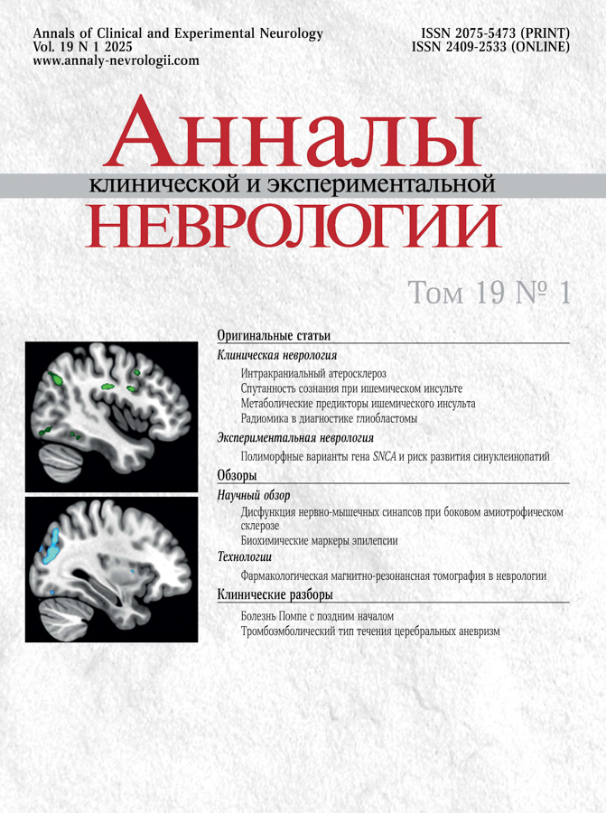
Технология фармакологической функциональной МРТ: потенциал использования в неврологии
- Авторы: Раскуражев А.А.1, Танашян М.М.1, Морозова С.Н.1, Кузнецова П.И.1, Аннушкин В.А.1, Мазур А.С.1, Панина А.А.1, Спрышков Н.Е.1, Пирадов М.А.1
-
Учреждения:
- Научный центр неврологии
- Выпуск: Том 19, № 1 (2025)
- Страницы: 68-76
- Раздел: Обзоры
- Статья получена: 17.01.2025
- Статья одобрена: 27.01.2025
- Статья опубликована: 03.04.2025
- URL: https://annaly-nevrologii.com/pathID/article/view/1267
- DOI: https://doi.org/10.17816/ACEN.1267
- ID: 1267
Цитировать
Аннотация
В обзоре представлены современные данные об одной из перспективных нейровизуализационных методик — фармакологической функциональной магнитно-резонансной томографии (фарм-фМРТ). Описаны технологии проведения фарм-фМРТ, варианты применения парадигмы в качестве триггера нейрональной активации зон интереса при изучении эффектов нейроактивных препаратов. Рассмотрены потенциальные возможности применения фарм-фМРТ при различных неврологических состояниях, таких как цереброваскулярные заболевания, эпилепсия, а также в отношении коррекции метаболических расстройств, когнитивных нарушений, болевого синдрома и др. Представлены ограничения применения фарм-фМРТ, возможные пути их преодоления при планировании и проведении исследований. Предложены перспективы применения фарм-фМРТ, которые позволят дать объективную оценку таргетного воздействия фармакологических агентов.
Полный текст
Введение
Сложности разработки и исследования лекарственных препаратов, влияющих на центральную нервную систему (ЦНС), подчёркивают необходимость поиска методологии и подходов, которые позволят осуществить качественный трансляционный скачок — от доклинических моделей к использованию и предикции клинического эффекта у пациентов [1]. Лабораторные in vitro и in vivo эксперименты способствуют описанию различных фармакологических свойств испытываемых молекул, однако данные, которые в ходе таких исследований указывают на эффективность препарата, следует (особенно для препаратов с нейроактивными характеристиками) интерпретировать с особой осторожностью в отношении человеческой популяции. На сегодняшний день не существует общеупотребимого референса при определении действия препаратов на ЦНС — будь то нейропротекция, антидепрессанты, нейролептики и др. [2]. Одной из перспективных методик может служить нейрофармакологическая функциональная магнитно-резонансная томография (фМРТ) головного мозга.
Технологии фМРТ, использующие относительный церебральный объём крови [3], сигнал, зависящий от уровня оксигенации крови (blood oxygen level dependent — так называемый BOLD-сигнал) [4], или Т1-взвешенную методику оценки мозгового кровотока [5], произвели революцию в картировании мозга [6]. Все указанные подходы основаны на сопряжении между нейрональной активностью, метаболизмом и гемодинамическими свойствами — параметрами, к которым чувствительны изменения интенсивности МР-сигнала. Несмотря на то что все упомянутые методики можно назвать функциональными, традиционно под термином «фМРТ» понимают оценку BOLD-сигнала.
Общеупотребимо в фМРТ-исследованиях использование некоторой парадигмы (например, зрительной стимуляции, движения пальцами, когнитивной задачи и др.) в качестве триггера нейрональной активации зон интереса (так называемая task-based fMRI). Впрочем, подобного эффекта можно достигнуть с помощью разного рода фармакологических агентов — как в качестве непосредственного стимула, так и в виде посредника, модулирующего ответ мозга на другую парадигму (например, когнитивную). Такую разновидность фМРТ в 1997 г. Y.C. Chen и соавт. обозначили как фармакологическую МРТ (фарм-фМРТ) [7]. В опытах на мышах они изучали регионарную селективность дофаминергических лигандов (однако идейно схожий подход был реализован и другими исследователями не позднее 1993 г. [8, 9]).
На ранних этапах клинических исследований методики фМРТ могут продемонстрировать функциональное воздействие фармакологического агента на ЦНС — причём в тех регионах мозга, которые этио- и/или патогенетически объяснимы с точки зрения биохимизма процессов [10]. Необходимо отдельно уточнить, что речь не идёт технически о маркерах таргетного действия (например, визуализация связывания фармпрепарата с соответствующим сайтом), а скорее о косвенном свидетельстве эффекта между фМРТ-ответом и биологическим правдоподобием действия агента. Установленные при фМРТ дозозависимые ассоциации могут стать ценными при планировании дальнейших этапов исследования или внедрении препарата в клиническую практику [11].
На более поздних фазах клинических испытаний данные фарм-фМРТ, вероятнее всего, будут полезны при попытке демонстрации нормализации изменённого в связи с заболеванием МР-сигнала (например, активации/деактивации определённых участков головного мозга в ответ на парадигму или изменение функциональной коннективности). Потенциально это можно рассматривать в качестве более объективной оценки модификации патологии ЦНС.
Фарм-фМРТ, помимо прочего, может быть использована с целью определения церебральных мишеней изучаемых фармакологических соединений, уточнения ожидаемых/непредвиденных механизмов действия, установления дозозависимых реакций, обеспечения валидных маркеров терапевтического ответа (в том числе в рамках клинических исследований) [12]. С момента выделения методики фарм-фМРТ на экспериментальных и/или клинических моделях был изучен достаточно широкий спектр нейроактивных молекул — как химических соединений (никотин, амфетамин и др.), так и терапевтических агентов (нейропептиды, холинергические, серотонинергические и глутаматергические препараты, каннабиноиды, опиоиды и др.) [14].
Применение фарм-фМРТ в качестве релевантной диагностической и исследовательской единицы (в том числе при разработке лекарственных средств) требует наличия определённых характеристик методики:
- данные фарм-фМРТ должны быть воспроизводимыми и изменяться под воздействием фармакологического агента;
- количественные характеристики фарм-фМРТ (с учётом оборудования) должны быть стандартизованы;
- особенности проведения и анализа фарм-фМРТ должны быть определены до начала исследования;
- для дальнейших исследований должна быть доступна имплементация выбранных методик фМРТ в нескольких центрах (например, при всех МР-исследованиях должны быть одинаковыми тип импульсных последовательностей, размеры вокселя, толщина среза, временнóе разрешение, угол отклонения, выбранные парадигмы);
- МРТ-лаборанты должны быть активно вовлечены в процесс проведения фМРТ (например, при обнаружении избыточных движений головой необходимо выполнение повторного сканирования);
- на всех этапах должен проводиться контроль качества (DICOM1 проверка следования протоколу, выявление артефактов и т. д.).
Одной из основных предпосылок изучения фарм-фМРТ является тот факт, что лекарственные препараты могут вызывать краткосрочные и долговременные изменения фМРТ-сигнала. Большое число опубликованных работ свидетельствует о том, что результаты фМРТ могут быть чувствительны как к краткой (после первой дозы), так и к долговременной (хронической, после многих приёмов) фармакотерапии. Например, в нескольких исследованиях показано, что фМРТ-ответ зоны амигдалы увеличивается на демонстрацию фотографий лиц с негативными эмоциями у пациентов с депрессией, в то время как приём антидепрессантов в клинически эффективной дозировке нормализует этот ответ [15, 16]. Другие группы препаратов, которые, как считается, могут индуцировать изменения фарм-фМРТ-сигналов, включают анальгетики, антипсихотики, блокаторы кальциевых каналов, ингибиторы циклооксигеназы-2, иммунотерапия.
Во многих случаях таргеты нейроактивных молекул известны из результатов ранее проведённых экспериментальных исследований (например, позитронно-эмиссионной томографии). При этом суммарный эффект фарм-фМРТ в ходе исследования можно подразделить на специфический (непосредственно связанный с рецепторной активацией) и общий, или неспецифический (связанный с побочными эффектами, которые могут влиять на интенсивность фМРТ-сигнала).
Фарм-фМРТ может идентифицировать единые точки приложения лекарственных средств. Схожие паттерны фМРТ-активации могут наблюдаться при использовании препаратов с одинаковыми показаниями к применению (например, болевой синдром), но при этом категорически отличных по механизму действия (например, нестероидные противовоспалительные препараты, опиодные анальгетики и др.). Более того, функциональный статус (фМРТ-характеристики) определённых структур мозга может служить предиктором терапевтического ответа на лечение — в частности, в случае прегабалина это показано для островка и нижней теменной дольки [17].
Описание технологии с современных позиций
Для получения BOLD-сигнала чаще всего используется эхо-планарная последовательность (градиентное эхо), чувствительная к локальным неоднородностям магнитного поля, которые могут быть вызваны, в частности, таким парамагнетиком, как дезоксигемоглобин, в присутствии которого сигнал снижается [5]. При возникновении возбуждения в определённой зоне головного мозга отмечается локальное усиление кровотока этой области. Это приводит к увеличению концентрации оксигемоглобина и снижению концентрации дезоксигемоглобина. В результате отмечается изменение (повышение) сигнала, которое регистрируется с помощью описанной последовательности. Таким образом, фМРТ позволяет регистрировать распределение нейрональной активности, о которой косвенно судят по изменению сигнала в зависимости от уровня оксигенации крови в сосудах головного мозга. В каждом вокселе регистрируются колебания этого сигнала в течение времени сканирования, которые затем оцениваются с помощью различных статистических инструментов с последующей возможностью группового представления данных, а также межгруппового и внутригруппового анализа (рис. 1).
Рис. 1. Результаты внутригруппового сравнения активации мозга при выполнении когнитивной парадигмы до и после лечения.
А — пациенты, получавшие сосудисто-метаболическую терапию в течение 10 дней (уменьшение активации в надкраевых и ангулярных извилинах, а также зрительной коре); В — пациенты, получавшие плацебо (уменьшение активации только в зрительной коре).
1 – аксиальная проекция; 2 — коронарная проекция; 3 — сагиттальная проекция.
Колебания сигнала происходят не только при выполнении какого-либо задания (task-based fMRI), но и спонтанно в состоянии покоя. В связи с внутренней нейрональной активностью регистрация подобных колебаний осуществляется при выполнении фМРТ покоя (rest fMRI) [18], т. е. сканирования пациента без предъявления ему стимулов любой модальности. Если колебания BOLD-сигнала между областями серого вещества похожи и коррелируют, то высока вероятность, что эти зоны функционально связаны. Исходя из этого постулата с помощью разных методов математического анализа были описаны многочисленные сети покоя мозга. Такой метод обладает рядом преимуществ перед фМРТ с парадигмами, в частности — возможностью проведения исследования даже в том случае, если пациент не способен понять или выполнить задание, а также отсутствием необходимости применять различную аппаратуру для предъявления стимулов, а следовательно, — меньшими трудоёмкостью и стоимостью. Тем не менее обработка таких данных в связи с наличием множества физиологических шумов более сложна и подвержена ошибкам.
Для фарм-фМРТ-исследований используются обе описанные методики [19, 20]. Введение лекарственного вещества или иного субстрата, потенциально способного изменять функциональную активность головного мозга, может воздействовать на BOLD-сигнал как на сосудистом, так и на нейрональном уровне, затрудняя в таком ключе интерпретацию результатов. Изменения сигнала, связанные с введением вещества в организм человека, невелики. Это оправдывает изучение его эффектов в рамках изменений сигнала при фМРТ с парадигмой, а именно, связанные с предъявляемым стимулом зоны активации у испытуемых с введением какого-либо вещества сравнивают с зонами активации у участников без введения или с использованием плацебо. Преимущества фМРТ покоя заключаются в том, что, помимо более простой методики проведения, она даёт возможность оценки влияния вводимого вещества на сетевом уровне, в том числе вдали от предполагаемой зоны максимальной концентрации рецепторов к веществу или ожидаемой области активации/деактивации [10].
Тем не менее для проведения валидного фарм-фМРТ-исследования дополнительно следует учитывать ряд нюансов, касающихся фармакокинетики и фармакодинамики вводимых в организм веществ, например, время до достижения максимальной концентрации вещества в крови, период полувыведения, накопительный эффект для выполнения исследования в момент максимального воздействия вещества на организм человека. Кроме того, следует учитывать и приём других веществ, которые могут взаимодействовать с изучаемым или сами по себе изменять функциональную активность головного мозга [14]. Исходя из этих данных рассчитывают время проведения исследования после введения в организм изучаемого вещества, а также время исследования в динамике после курса терапии.
Использование метода фарм-фМРТ в различных сферах неврологии
Цереброваскулярные заболевания
Имеющиеся данные об исследовании методики фарм-фМРТ при нарушениях мозгового кровообращения ограничены прежде всего хронической цереброваскулярной патологией и особой группой лекарственных средств — нейропротекторами. Одними из пилотных в этом направлении стали исследования Научного центра неврологии. Так, в работе 2010 г. курсовое применение одного из препаратов с заявленным нейропротективным действием было ассоциировано с расширением имеющихся зон и/или появлением новых зон активации, преимущественно в теменно-затылочной области, что сочеталось с улучшением выполнения основных когнитивных тестов [21]. Напротив, проведённое годом позже исследование нейропептидного препарата продемонстрировало уменьшение зон активации (особенно в височных и лобных долях) в ответ на разработанную в Научном центре неврологии оригинальную когнитивную парадигму [22]. Непрямое фарм-фМРТ-сравнение нескольких потенциальных нейропротекторных препаратов позволило предположить основные механизмы таргетного действия и фарм-фМРТ-паттерны: цереброактивирующее действие, улучшение микроциркуляции, уменьшение энергетических затрат мозга, нейрометаболический эффект [23].
Достаточно интересны результаты фарм-фМРТ-исследования отечественного нейропротекторного препарата, в которых после лечения отмечалось уменьшение зон, необходимых для выполнения когнитивной задачи (в надкраевой и ангулярной извилинах), улучшение управляющих функций мозга, связанных с процессингом языковой информации (усиление связи между левой дорсолатеральной префронтальной корой с отделами верхней височной извилины). Клинически эти нейровизуализационные изменения проявлялись повышением функциональной активности и оптимизацией исполнительных функций, что является важным патогенетическим эффектом для пациентов с сосудистой патологией мозга [24].
Болевые синдромы
Ряд исследований посвящён изучению анальгетических возможностей опиоидных препаратов на активность структур головного мозга [25, 26]. Опиоидный анальгетик налбуфин, повышал интенсивность BOLD-сигнала в 60 областях головного мозга при одновременном снижении его в 9 областях, включая среднюю лобную кору, нижнюю орбитофронтальную кору, постцентральную теменную кору, верхнюю височную извилину и мозжечок. Однако при введении налоксона паттерн изменённой активации существенно преобразился: повышение интенсивности BOLD-сигнала было отмечено лишь в 14 зонах, снижение — в 3. Низкие дозы налоксона значительно блокировали активность налбуфина в верхней медиальной и средней лобной коре, постцентральной теменной коре, затылочной коре (роландовой борозде), хвостатом ядре, мосту (главном сенсорном ядре тройничного нерва) и мозжечке.
Отдельное место в противоболевой терапии на сегодняшний день занимают антидепрессанты и антиконвульсанты, демонстрирующие мультимодальные возможности в контроле над хроническим болевым синдромом. A.E. Edes и соавт. исследовали роль внутривенного введения циталопрама/плацебо у 27 здоровых добровольцев и 6 пациентов с мигренью без ауры на активность передней поясной коры как основной структуры, участвующей в нисходящей модуляции и эмоциональном аспекте боли [27]. Выявлена значимая разница во временнóм паттерне активации передней поясной коры между здоровыми добровольцами контрольной группы и пациентами с мигренью без ауры во время даже небольшого повышения уровня серотонина на фоне приёма циталопрама.
Эпилепсия
Достаточно широко представлены исследования фарм-фМРТ в контексте эпилепсии. С учётом разнообразия доступных противоэпилептических препаратов (ПЭП) и неоднородности эпилептических синдромов с точки зрения задействованных нейронных сетей существует необходимость разработки биомаркеров по данным фМРТ для раннего определения эффективности лечения и вероятности побочных эффектов [10]. Так, увеличение дозировки вальпроевой кислоты у пациентов с юношеской миоклонической эпилепсией, по данным фарм-фМРТ, было ассоциировано с ослаблением аномальной коактивации двигательной коры с когнитивными сетями во время исследования рабочей памяти [28]. Применение другого ПЭП — леветирацетама у пациентов с височной эпилепсией, согласно исследованиям фарм-фМРТ, сопровождалось восстановлением нормального паттерна активации [29]: в частности, на фоне приёма препарата отмечено усиление дезактивации в ответ на когнитивную парадигму в поражённой височной доле, причём подтверждён дозозависимый эффект.
Результаты исследований топирамата демонстрируют потенциальную роль фарм-фМРТ в уточнении церебральных механизмов нежелательных явлений нейрофармакологических средств. На фоне приёма топирамата (как при эпилепсии, так и у пациентов с мигренью и здоровых добровольцев) применение арсенала фарм-фМРТ позволило выявить паттерн сниженной активации в языко-зависимых участках мозга (нижняя и средняя лобные извилины, верхняя височная извилина доминантного полушария) [30–32], а также отсутствие феномена деактивации парадигм-независимых зон, включая сеть пассивного режима работы [33, 34].
Цереброметаболическое здоровье
В контексте концепции цереброметаболического здоровья, охватывающей большой пласт синдемии неврологических и метаболических заболеваний, актуально изучение влияния различных препаратов с целью коррекции тех или иных симптомов [35].
На сегодняшний день исследователи располагают несколькими модальностями для изучения пищевого поведения, основным из них является нейрокогнитивное тестирование с помощью различных опросников. С появлением фМРТ стало возможно в режиме реального времени оценивать изменение активации структур головного мозга в ответ на различные стимулы (например, с помощью зрительной пищевой парадигмы). Основные зоны, исследуемые у пациентов с ожирением, — «система награды», составными частями которой являются префронтальная кора, островковая доля, поясная извилина и лимбическая система. На базе Научного центра неврологии разработана простая и воспроизводимая зрительная фМРТ-парадигма для оценки системы контроля пищевого поведения [36], которая в дальнейшем использовалась в исследованиях, в том числе фарм-фМРТ-паттернов. Так, на фоне приёма сибутрамина (средство для лечения ожирения центрального действия, механизм действия которого обусловлен селективным ингибированием обратного захвата серотонина и норадреналина) показан различный паттерн изменения сигнала в ответ на пищевую парадигму у пациентов с ожирением по сравнению со здоровыми добровольцами. Наиболее существенные изменения функциональной активности отмечены в затылочных долях, островке, средней и верхней лобных извилинах. Интересным является тот факт, что до назначения фармакотерапии у пациентов с ожирением по сравнению с контрольной группой (здоровые добровольцы) прежде всего обращала на себя внимание чрезмерная активность затылочных долей, что косвенно свидетельствует о более значимой эмоциональной реакции на демонстрацию высококалорийной пищи у людей с избыточной массой тела [37].
O. Farr и соавт. с участием 20 пациентов с сахарным диабетом 2-го типа показали влияние лираглутида (аналог человеческого глюкагон-подобного пептида 1) на активацию зон головного мозга (дорсолатеральная префронтальная кора, средний мозг, таламическая область) в ответ на пищевые стимулы [38]. В исследовании H. Cheng и соавт. продемонстрированы мультимодальные эффекты лираглутида на когнитивные функции в виде повышения активации в области гиппокампа, что расширяет возможности применения препаратов данной группы у пациентов с сахарным диабетом 2-го типа и ожирением [39].
В последние десятилетия значительно возрос интерес к изучению влияния различных пищевых веществ на мозг и поведение человека. Сахар и искусственные подсластители широко используются в современной диетологии. фМРТ головного мозга позволяет изучить эти механизмы с возможностью оценки динамики нейрональной активности в ответ на потребление пищевых веществ. Известно, что глюкоза активирует системы вознаграждения в мозге (например, дофаминовую систему), что связано с приятными ощущениями и мотивацией. Понимание того, как быстроусвояемые углеводы, в частности сахароза, активируют различные зоны мозга у здоровых людей, может дать ключ к пониманию механизмов переедания и зависимости.
Сахарозаменители (аспартам, сукралоза, стевия, эритрит) предлагаются как более здоровая альтернатива сахарозе и фруктозе. Однако их влияние на мозговую активность и поведение человека остаётся предметом дискуссии. Некоторые исследования показывают, что сахарозаменители могут не активировать системы вознаграждения как углеводы, что может влиять на чувство насыщения и потребление пищи в дальнейшем. В контексте ожирения фМРТ используется для оценки функциональной нейрональной активности, участвующей в регуляции энергетического обмена и метаболизма. В Научном центре неврологии получены пилотные результаты сравнения эффектов сахарозы и сахарозаменителя с применением фМРТ, которые показали различия в активации в области дополнительной моторной и дорсолатеральной префронтальной коры среди здоровых добровольцев (рис. 2).
Рис. 2. Внутригрупповое сравнение активации головного мозга здоровых испытуемых при визуализации пищевой парадигмы (изображения аппетитной и неаппетитной еды) после приёма сахара и сахарозаменителя. На срезах головного мозга представлены зоны с отличающейся активацией. После приёма сахара отмечается бóльшая активация в дополнительной моторной и дорсолатеральной префронтальной коре с обеих сторон.
А — аксиальная проекция; В — коронарная проекция.
Когнитивные нарушения
Фарм-фМРТ может явиться перспективным инструментом для идентификации таргетов препаратов, используемых для коррекции когнитивных нарушений. Убедительно продемонстрирован дифференцированный эффект холинергической терапии (галантамин) в зависимости от целевой когорты пациентов: умеренные когнитивные нарушения (активация задней поясной извилины, левой нижней теменной и передней височной долей) или болезнь Альцгеймера (двусторонняя активация гиппокампа) [40]. Подобные изменения в реакции на холинергическую нагрузку могут отражать исходную разницу в функциональном состоянии холинергической системы между обеими группами, что согласуется с клиническими исследованиями. Более того, выявлены различия в паттернах активации при однократном и длительном приёме препарата, что подчёркивает важность изучения фарм-фМРТ в качестве динамической методики.
Основные приложения технологии фарм-фМРТ в неврологии:
- исследование «классических» нейропротекторов у пациентов с цереброваскулярными заболеваниями;
- исследование антидепрессантов у пациентов неврологического профиля (постинсультная депрессия, хронический болевой синдром, нейродегенерация и др.);
- оценка resting state у пациентов с эпилепсией в зависимости от фармакокинетики/динамики ПЭП;
- лечение острой/хронической боли;
- оценка фМРТ коррелятов нейропластичности у пациентов после инсульта;
- холинергическая терапия у пациентов с когнитивными расстройствами (сосудистая деменция, болезнь Альцгеймера, болезнь Паркинсона и др.);
- дофаминергическая терапия у пациентов с паркинсонизмом;
- пациенты с рассеянным склерозом на фоне пульс-терапии кортикостероидами.
Технологические сложности методики фарм-фМРТ и возможные пути преодоления
Фарм-фМРТ является сложной методикой, применение которой сопряжено с высоким уровнем материально-технических затрат, а интерпретация результатов требует осторожности и взвешенного подхода.
Приведём далеко не полный список существующих ограничений этой технологии:
- отсутствие оптимального набора настроек для сбора и процессинга МР-изображений;
- ограничения обобщённой линейной модели как основного подхода статистического анализа фМРТ-данных;
- отсутствие стандартизированных парадигм под конкретные цели исследования;
- существующие стандарты представления данных фарм-фМРТ экспериментов недостаточны для адекватной оценки и интерпретации;
- предвзятость в отношении проведения и публикации валидационных (повторных) фМРТ-исследований;
- использование BOLD-сигнала в качестве прокси-индикатора зависит от исходного уровня нейроваскулярного сопряжения, а модуляция последнего под воздействием фармакологических агентов зачастую сложно прогнозируема;
- для большинства исследований — малая численность выборки и значительная её гетерогенность;
- высокая меж- и внутрииндивидуальная вариабельность фМРТ-сигнала [14, 41].
Понимание ограничений технологии, а также вышеописанных особенностей методики фарм-фМРТ может позволить (по крайней мере, частично) модифицировать методологию исследования для получения воспроизводимых и значимых результатов. Так, выбор нейроактивных молекул для эксперимента должен базироваться не только на клинической целесообразности, но и особенностях фармакокинетики/-динамики и таргетного взаимодействия; при этом следует обязательно учитывать время и продолжительность ожидаемого эффекта, что необходимо для построения правильного дизайна работы. Поскольку изменения BOLD-сигнала, наблюдаемые в ходе фМРТ, могут быть обусловлены системными эффектами (частотой сердечных сокращений, уровнем сатурации крови и др.), желательно учитывать эти рутинные показатели при статистическом анализе [42]. Нормализация исходных различий цереброваскулярной реактивности между пациентами и в контексте применения плацебо/активного препарата является желательной для преодоления ограничений, заложенных в методику оценки BOLD-сигнала. Для выполнения этой задачи возможно использование оценки базового уровня церебральной перфузии (с помощью метода меченых артериальных спинов) [43], измерение скорости церебрального метаболизма потребления кислорода [44].
При выполнении фарм-фМРТ с различными парадигмами большое значение имеет проведение отдельных (отстоящих друг от друга на дни/недели) сканирований с использованием плацебо [45]. Помимо этого, исследование фармакологических агентов с преимущественно субъективными эффектами (например, вызывающими сонливость или, наоборот, прилив сил, модулирующими настроение и т. п.) следует дополнить психометрическими тестами в заранее оговорённые временны́е промежутки в течение сканирований и/или между ними (в случае предполагаемого долгосрочного эффекта) [46]. В некоторых случаях методике фарм-фМРТ с парадигмой следует предпочесть (или дополнить) фМРТ покоя, поскольку последняя позволяет провести анализ функциональной коннективности и выявить потенциально более устойчивые маркеры ответа на терапию [47].
Заключение
Фарм-фМРТ головного мозга, являясь одной из множества подвидов ангионейровизуализационных методик, обладает значительными перспективами для изучения в области нейронаук. Этот метод позволяет при правильном дизайне исследования обеспечить in vivo объективную оценку таргетного воздействия фармакологического агента. Это принципиально с нескольких точек зрения:
- персонификация назначаемой терапии (например, в случае коррекции противоэпилептической терапии);
- подтверждение и/или открытие новых механизмов действия нейроактивных препаратов (особенно актуально для нейропротекторов);
- сокращение сроков разработки новых лекарственных средств благодаря прямой визуализации наличия/отсутствия церебрального эффекта;
- уточнение генеза нежелательных явлений нейроактивных препаратов;
- расширение арсенала фундаментальных наук и возможностей изучения специфических рецепторов.
Совместные с фармакологической промышленностью разработки в рамках лабораторий фарм-фМРТ, оснащённых передовым оборудованием, могут обеспечить конкурентное преимущество отечественных разработок и ускоренную трансляцию результатов экспериментальных нейронаук в клиническую практику. Вместе с тем методика обладает значительным спектром ограничений, преодоление которых является такой же полноценной и важной задачей, как и непосредственное изучение проблемы.
1 DICOM (Digital Imaging and Communications in Medicine) — медицинский отраслевой стандарт создания, хранения, передачи и визуализации цифровых медицинских изображений и документов обследованных пациентов.
Об авторах
Антон Алексеевич Раскуражев
Научный центр неврологии
Автор, ответственный за переписку.
Email: raskurazhev@neurology.ru
ORCID iD: 0000-0003-0522-767X
канд. мед. наук, врач-невролог, с. н. с. 1-го неврологического отделения, рук. лаб. нейрофармакологической функциональной МРТ Института клинической и профилактической неврологии
Россия, 125367, Москва, Волоколамское шоссе, д. 80Маринэ Мовсесовна Танашян
Научный центр неврологии
Email: raskurazhev@neurology.ru
ORCID iD: 0000-0002-5883-8119
д-р мед. наук, профессор, член-корреспондент РАН, заместитель директора по научной работе, руководитель 1-го неврологического отделения Института клинической и профилактической неврологии
Россия, 125367, Москва, Волоколамское шоссе, д. 80Софья Николаевна Морозова
Научный центр неврологии
Email: raskurazhev@neurology.ru
ORCID iD: 0000-0002-9093-344X
канд. мед. наук, н. с. отдела лучевой диагностики Института клинической и профилактической неврологии
Россия, 125367, Москва, Волоколамское шоссе, д. 80Полина Игоревна Кузнецова
Научный центр неврологии
Email: raskurazhev@neurology.ru
ORCID iD: 0000-0002-4626-6520
канд. мед. наук, врач-невролог, н. с. 1-го неврологического отделения Института клинической и профилактической неврологии
Россия, 125367, Москва, Волоколамское шоссе, д. 80Владислав Александрович Аннушкин
Научный центр неврологии
Email: raskurazhev@neurology.ru
ORCID iD: 0000-0002-9120-2550
канд. мед. наук, врач-невролог 1-го неврологического отделения Института клинической и профилактической неврологии
Россия, 125367, Москва, Волоколамское шоссе, д. 80Андрей Сергеевич Мазур
Научный центр неврологии
Email: raskurazhev@neurology.ru
ORCID iD: 0000-0001-8960-721X
аспирант 1-го неврологического отделения Института клинической и профилактической неврологии
Россия, 125367, Москва, Волоколамское шоссе, д. 80Анастасия Андреевна Панина
Научный центр неврологии
Email: raskurazhev@neurology.ru
ORCID iD: 0000-0002-8652-2947
аспирантка 1-го неврологического отделения Института клинической и профилактической неврологии
Россия, 125367, Москва, Волоколамское шоссе, д. 80Никита Евгеньевич Спрышков
Научный центр неврологии
Email: raskurazhev@neurology.ru
ORCID iD: 0000-0002-2934-5462
аспирант 1-го неврологического отделения Института клинической и профилактической неврологии
Россия, 125367, Москва, Волоколамское шоссе, д. 80Михаил Александрович Пирадов
Научный центр неврологии
Email: raskurazhev@neurology.ru
ORCID iD: 0000-0002-6338-0392
д-р мед. наук, профессор, академик РАН, директор
Россия, 125367, Москва, Волоколамское шоссе, д. 80Список литературы
- Dawson GR, Craig KJ, Dourish CT. Validation of experimental medicine methods in psychiatry: the P1vital approach and experience. Biochem Pharmacol. 2011;81(12):1435–1441. doi: 10.1016/j.bcp.2011.03.013
- Conn PJ, Roth BL. Opportunities and challenges of psychiatric drug discovery: roles for scientists in academic, industry, and government settings. Neuropsychopharmacology. 2008;33(9):2048–2060. doi: 10.1038/sj.npp.1301638
- Bandettini PA, Wong EC, Hinks RS, et al. Time course EPI of human brain function during task activation. Magn. Reson. Med. 1992;25(2):390–397. doi: 10.1002/mrm.1910250220
- Ogawa S, Tank DW, Menon R, et al. Intrinsic signal changes accompanying sensory stimulation: functional brain mapping with magnetic resonance imaging. Proc Natl Acad Sci USA. 1992;89(13):5951–5955. doi: 10.1073/pnas.89.13.5951
- Kwong KK, Belliveau JW, Chesler DA, et al. Dynamic magnetic resonance imaging of human brain activity during primary sensory stimulation. Proc Natl Acad Sci U S A. 1992;89(12):5675–5679. doi: 10.1073/pnas.89.12.5675
- Jenkins BG. Pharmacologic magnetic resonance imaging (phMRI): imaging drug action in the brain. Neuroimage. 2012;62(2):1072–1085. doi: 10.1016/j.neuroimage.2012.03.075
- Chen YC, Galpern WR, Brownell AL, et al. Detection of dopaminergic neurotransmitter activity using pharmacologic MRI: correlation with PET, microdialysis, and behavioral data. Magn Reson Med. 1997;38(3):389–398. doi: 10.1002/mrm.1910380306
- Silva AC, Zhang W, Williams DS, Koretsky AP. Multi-slice MRI of rat brain perfusion during amphetamine stimulation using arterial spin labeling. Magn Reson Med. 1995;33(2):209–214. doi: 10.1002/mrm.1910330210
- Cuenod CA, Chang MCJ, Arai T, et al. Local brain response to cholinergic receptor stimulation detected by MRI. Proc Int Soc Magn Reson Med. 1993:S3;1387.
- Wandschneider B, Koepp MJ. Pharmaco fMRI: determining the functional anatomy of the effects of medication. Neuroimage Clin. 2016;12:691–697. doi: 10.1016/j.nicl.2016.10.002
- Upadhyay J, Anderson J, Baumgartner R, et al. Modulation of CNS pain circuitry by intravenous and sublingual doses of buprenorphine. Neuroimage. 2012;59(4):3762–3773. doi: 10.1016/j.neuroimage.2011.11.034
- Fanny M, Manuel T, Daniel HW, et al. Pharmacological manipulation of neurotransmitter activity induces disparate effects on cerebral blood flow and resting-state fluctuations. Imaging Neuroscience. 2024;2:1–18. doi: 10.1162/imag _a_00370
- Jenkins BG. Pharmacologic magnetic resonance imaging (phMRI): imaging drug action in the brain. Neuroimage. 2012;62(2):1072–1085. doi: 10.1016/j.neuroimage.2012.03.075
- Carmichael O, Schwarz AJ, Chatham CH, et al. The role of fMRI in drug development. Drug Discov Today. 2018;23(2):333–348. doi: 10.1016/j.drudis.2017.11.012
- Delaveau P, Jabourian M, Lemogne C, et al. Brain effects of antidepressants in major depression: a meta-analysis of emotional processing studies. J Affect Disord. 2011;130(1-2):66–74. doi: 10.1016/j.jad.2010.09.032
- van Wingen GA, Tendolkar I, Urner M, et al. Short-term antidepressant administration reduces default mode and task-positive network connectivity in healthy individuals during rest. Neuroimage. 2014;88:47–53. doi: 10.1016/j.neuroimage.2013.11.022
- Harris RE, Napadow V, Huggins JP, et al. Pregabalin rectifies aberrant brain chemistry, connectivity, and functional response in chronic pain patients. Anesthesiology. 2013;119(6):1453–1464. doi: 10.1097/ALN.0000000000000017
- Biswal B, Yetkin FZ, Haughton VM, Hyde JS. Functional connectivity in the motor cortex of resting human brain using echo-planar MRI. Magn Reson Med. 1995;34(4):537–541. doi: 10.1002/mrm.1910340409
- Gaebler AJ, Fakour N, Stöhr F, et al. Functional connectivity signatures of NMDAR dysfunction in schizophrenia-integrating findings from imaging genetics and pharmaco-fMRI. Transl. Psychiatry. 2023;13(1):59. doi: 10.1038/s41398-023-02344-2
- Berginström N, Nordström P, Ekman U, et al. Pharmaco-fMRI in patients with traumatic brain injury: a randomized controlled trial with the monoaminergic stabilizer (-)-OSU6162. J Head Trauma Rehabil. 2019;34(3):189–198. doi: 10.1097/HTR.0000000000000440
- Танашян М.М., Лагода О.В., Федин П.А. и др. Современные подходы к лечению больных с хроническими сосудистыми заболеваниями головного мозга. Нервные болезни. 2010;(4):19–22. Tanashyan MM, Lagoda OV, Fedin PA, et al. Modern approaches to the treatment of patients with chronic vascular diseases of the brain. Nervnyye bolezni. 2010;(4):19–22.
- Танашян М.М., Бархатов Д.Ю., Глотова Н.А. и др. Эффективность нейропротекции у больных с хроническими цереброваскулярными заболеваниями. Вестник Российской военно-медицинской академии. 2011;3(35):181–187. Tanashyan MM, Barkhatov DYu, Glotova NA, et al. The effectiveness of neuroprotection in patients with chronic cerebrovascular diseases. Bulletin of the Russian Military Medical Academy, 2011;3(35):181–187.
- Танашян М.М., Коновалов Р.Н., Лагода О.В. Новые подходы к коррекции когнитивных нарушений при цереброваскулярных заболеваниях. Анналы клинической и экспериментальной неврологии. 2018;12(3):30–39. doi: 10.25692/ACEN.2018.3.4 Tanashyan MM, Konovalov RN, Lagoda OV. New approaches to correction of cognitive impairments in cerebrovascular diseases. Annals of clinical and experimental Neurology. 2018;12(3):30–39. doi: 10.25692/ACEN.2018.3.4
- Tanashyan M, Morozova S, Raskurazhev A, Kuznetsova P. A prospective randomized, double-blind placebo-controlled study to evaluate the effectiveness of neuroprotective therapy using functional brain MRI in patients with post-covid chronic fatigue syndrome. Biomed Pharmacother. 2023;168:115723. doi: 10.1016/j.biopha.2023.115723
- Becerra L, Harter K, Gonzalez RG, Borsook D. Functional magnetic resonance imaging measures of the effects of morphine on central nervous system circuitry in opioid-naive healthy volunteers. Anesth Analg. 2006;103(1):208–216. doi: 10.1213/01.ane.0000221457.71536.e0
- Gear R, Becerra L, Upadhyay J, et al. Pain facilitation brain regions activated by nalbuphine are revealed by pharmacological fMRI. PLoS One. 2013;8(1):e50169. doi: 10.1371/journal.pone.0050169
- Edes AE, McKie S, Szabo E, et al. Increased activation of the pregenual anterior cingulate cortex to citalopram challenge in migraine: an fMRI study. BMC Neurol. 2019;19(1):237. doi: 10.1186/s12883-019-1478-0
- Vollmar C, O’Muircheartaigh J, Symms MR, et al. Altered microstructural connectivity in juvenile myoclonic epilepsy: the missing link. Neurology. 2012;78(20):1555–1559. doi: 10.1212/WNL.0b013e3182563b44
- Wandschneider B, Stretton J, Sidhu M, et al. Levetiracetam reduces abnormal network activations in temporal lobe epilepsy. Neurology. 2014;83(17):1508–1512. doi: 10.1212/WNL.0000000000000910
- De Ciantis A, Muti M, Piccolini C, et al. A functional MRI study of language disturbances in subjects with migraine headache during treatment with topiramate. Neurol Sci. 2008;29(Suppl 1):S141–143. doi: 10.1007/s10072-008-0906-5
- Jansen JF, Aldenkamp AP, Marian Majoie HJ, et al. Functional MRI reveals declined prefrontal cortex activation in patients with epilepsy on topiramate therapy. Epilepsy Behav. 2006;9(1):181–185. doi: 10.1016/j.yebeh.2006.05.004
- Szaflarski JP, Allendorfer JB. Topiramate and its effect on fMRI of language in patients with right or left temporal lobe epilepsy. Epilepsy Behav. 2012;24(1):74–80. doi: 10.1016/j.yebeh.2012.02.022
- Tang Y, Xia W, Yu X, et al. Altered cerebral activity associated with topiramate and its withdrawal in patients with epilepsy with language impairment: an fMRI study using the verb generation task. Epilepsy Behav. 2016;59:98–104. doi: 10.1016/j.yebeh.2016.03.013
- Yasuda CL, Centeno M, Vollmar C, et al. The effect of topiramate on cognitive fMRI. Epilepsy Res. 2013;105(1-2):250-255. doi: 10.1016/j.eplepsyres.2012.12.007
- Танашян М.М., Антонова К.В. Цереброметаболическое здоровье. В кн.: Управление метаболическим здоровьем. М.; 2025;II:117–148. Tanashyan MM, Antonova KV. Cerebrometabolic health. In: Management of metabolic health. Moscow; 2025;II:119–148. (In Russ.)
- Кремнева Е.И., Суслин А.С., Говорин A.Н. и др. фМРТ-картирование алиментарных функциональных зон головного мозга. Анналы клинической и экспериментальной неврологии. 2015;9(1):32–36. doi: 10.17816/psaic156 Kremneva EI, Suslin AS, Govorin AN, et al. Mapping of the brain regions responsible for eating behavior regulation with functional MRI. Annals of clinical and experimental neurology. 2015;9(1):32–36. doi: 10.17816/psaic156
- Кузнецова П.И., Романцова Т.И., Логвинова О.В. и др. Функциональная МР-томография головного мозга на фоне медикаментозной коррекции ожирения. Ожирение и метаболизм. 2022;19(1):74–82. doi: 10.14341/omet12810 Kuznetsova PI, Romantsova TI, Logvinova OV, et al. Functional brain MRI in the setting of drug correction of obesity. Obesity and metabolism. 2022;19(1):74–82. doi: 10.14341/omet12810
- Farr OM, Tsoukas MA, Triantafyllou G, et al. Short-term administration of the GLP-1 analog liraglutide decreases circulating leptin and increases GIP levels and these changes are associated with alterations in CNS responses to food cues: a randomized, placebo-controlled, crossover study. Metabolism. 2016;65(7):945–953. doi: 10.1016/j.metabol.2016.03.009
- Cheng H, Zhang Z, Zhang B, et al. enhancement of impaired olfactory neural activation and cognitive capacity by liraglutide, but not dapagliflozin or acarbose, in patients with type 2 diabetes: a 16-week randomized parallel comparative study. Diabetes Care. 2022;45(5):1201–1210. doi: 10.2337/dc21-2064
- Goekoop R, Scheltens P, Barkhof F, et al. Cholinergic challenge in Alzheimer patients and mild cognitive impairment differentially affects hippocampal activation — a pharmacological fMRI study. Brain. 2006;129 (Pt 1):141–157. doi: 10.1093/brain/awh671
- Bourke JH, Wall MB. phMRI: methodological considerations for mitigating potential confounding factors. Front Neurosci. 2015;9:167. doi: 10.3389/fnins.2015.00167
- Glover GH, Li TQ, Ress D. Image-based method for retrospective correction of physiological motion effects in fMRI: RETROICOR. Magn Reson Med. 2000;44(1):162–167. doi: 10.1002/1522-2594(200007)44:1<162::aid-mrm23>3.0.co;2-e
- Murphy SE, Mackay CE. Using MRI to measure drug action: caveats and new directions. J Psychopharmacol. 2011;25(9):1168–1174. doi: 10.1177/0269881110372547
- Pattinson KT, Rogers R, Mayhew SD, et al. Pharmacological FMRI: measuring opioid effects on the BOLD response to hypercapnia. J Cereb. Blood Flow Metab. 2007;27(2):414–423. doi: 10.1038/sj.jcbfm.9600347
- Deakin JF, Lees J, McKie S, et al. Glutamate and the neural basis of the subjective effects of ketamine: a pharmaco-magnetic resonance imaging study. Arch Gen Psychiatry. 2008;65(2):154–164. doi: 10.1001/archgenpsychiatry.2007.37
- Anderson IM, Clark L, Elliott R, et al. 5-HT(2C) receptor activation by m-chlorophenylpiperazine detected in humans with fMRI. Neuroreport. 2002;13(12):1547–1551. doi: 10.1097/00001756-200208270-00012
- Cole DM, Smith SM, Beckmann CF. Advances and pitfalls in the analysis and interpretation of resting-state FMRI data. Front Syst Neurosci. 2010;4:8. doi: 10.3389/fnsys.2010.00008


