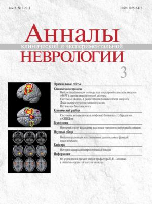
The fMRI study of 7 healthy volunteers was performed to evaluate the neuron networks of sensorimotor system during active and passive movements of the left and right index fingers. While performing all the tasks, the predominant activation of primary sensorimotor, premotor, supplementary motor areas, secondary sensory zones were shown on the contralateral side, as well as ipsilateral cerebellar activation. It was demonstrated that brain activation areas in both types of paradigm were corresponding, as well as the activation clusters size, amplitude and voxel coordinates, with the maximum values in the primary motor and sensory cortex. These results allow recommending the paradigm of passive index finger movements in given rate for the evaluation of sensorimotor system in patients with movement disorders.
Functional MRI study: passive motor paradigm in the assessment of sensorimotor system
- Authors: Dobrynina L.A.1, Kremneva E.I.1, Konovalov R.N.1, Kadykov A.S.1
-
Affiliations:
- Research Center of Neurology
- Issue: Vol 5, No 3 (2011)
- Pages: 11-19
- Section: Original articles
- Submitted: 03.02.2017
- Published: 13.02.2017
- URL: https://annaly-nevrologii.com/journal/pathID/article/view/302
- DOI: https://doi.org/10.17816/psaic302
- ID: 302
Cite item
Full Text
Abstract
About the authors
Larisa A. Dobrynina
Research Center of Neurology
Email: dobrla@mail.ru
ORCID iD: 0000-0001-9929-2725
D. Sci. (Med.), Head, 3rd Neurology department
Russian Federation, MoscowElena I. Kremneva
Research Center of Neurology
Email: dobrla@mail.ru
ORCID iD: 0000-0001-9396-6063
Cand. Sci. (Med.), senior researcher, Radiology department
Russian Federation, 125367, Russia, Moscow, Volokolamskoye shosse, 80Rodion N. Konovalov
Research Center of Neurology
Email: dobrla@mail.ru
ORCID iD: 0000-0001-5539-245X
Cand. Sci. (Med.), senior researcher, Neuroradiology department
Russian Federation, 125367 Moscow, Volokolamskoye shosse, 80Albert S. Kadykov
Research Center of Neurology
Author for correspondence.
Email: dobrla@mail.ru
ORCID iD: 0000-0001-7491-7215
D. Sci. (Med.), Professor, senior researcher, 3rd Neurological department
Russian Federation, MoscowReferences
- Arthurs O., Boniface S.How well do we understand the neural origins of the fMRI BOLD signal? Trends Neurosci. 2002; 25: 27–31.
- Baehr M., Frotscher M. Duus’ topical diagnosis in neurology: anatomy, physiology, signs, symptoms. Thieme, 4th 2005.
- Baron J.C., Cohen L.G., Cramer S.C. et al. Neuroimaging in stroke recovery: a position paper from the First International Workshop on Neuroimaging and Stroke Recovery. Cerebrovasc. Dis. 2004; 18: 260–267.
- Butefisch C.M., Kleiser R., Korber B. et al. Recruitment of contralesional motor cortex in stroke patients with recovery of hand function. Neurology 2005; 64: 1067–1069.
- Calautti C. and Baron J.-K. Functional neuroimaging studies of motor recovery after stroke in adults. Stroke 2003; 34: 1553–1566.
- Carey L.M., Abbot D.F., Egan G.F. et al. Evolution of brain activation with good and poor motor recovery after stroke. Neurorehabil. Neural. Repair. 2006; 20: 24–41.
- Casey K.L., Minoshima S., Morrow T.J., Koeppe R.A. Comparison of human cerebral activation pattern during cutaneous warmth, heat pain, and deep cold pain. J. Neurophysiol. 1996; 76: 571–581.
- Colebatch J.G., Deiber M.P., Passingham R.E. et al. Regional cerebral blood flow during voluntary arm and hand movements in human subjects. J. Neurophysiol. 1991; 65: 1392–1401.
- Cramer S.C., Nelles G., Benson R.R. et al. A functional MRI study of subjects recovered from hemiparetic stroke. Stroke 1997; 28: 2518–2527.
- Cramer S.C., Nelles G., Schaechter J.D. et al. A functional MRI study of three motor tasks in the evaluation of stroke recovery. Neurorehabil Neural. Repair 2001; 15: 1–8.
- Friston K.J., Holmes A.P., Worsley K.J. et al. Statistical parametric maps in functional imaging: A general linear approach. Human Brain Mapping 1995; 2 (4): 189–210.
- Goldring S., Ratcheson R. Human motor cortex: sensory input data from single neuron recordings. Science 1972; 175: 1493–1495.
- Ibanez V., Deiber M.P., Sadato N. et al. Effects of stimulas rate on regional cerebral blood flow after median nerve stimulation. Brain 1995; 118: 1339–1351.
- Kim Y.H., You S.H., Kwon Y.H. et al. Longitudinal fMRI study for locomotor recovery in patients with stroke. Neurology 2006; 67: 330–333.
- Kocak M., Ulmer J.L., Ugurel M.S. et al. Motor Homunculus: Passive mapping in healthy volunteers by using functional MR Imaging – initial results. Radiology 2009; 251: 485–492.
- Lancaster J.L., Woldorff M.G., Parsons L.M. et al. Automated Talairach atlas labels for functional brain mapping. Hum. Brain Mapp. 2000; 10: 120–131.
- Maldjian J.A., Laurienti P.J., Kraft R.A., Burdette J.H. An automated method for neuroanatomic and cytoarchitectonic atlas-based interrogation of fMRI data sets. Neuroimage 2003; 19: 1233–1239.
- Mima T., Sadato N., Yazawa S. et al. Brain structures related to active and passive finger movements in man. Brain 1999; 122: 1989–1997.
- Nudo R.J. Functional and structural plasticity in motor cortex: implications for stroke recovery. Phys. Med. Rehabil. Clin. N. Am. 2003; 14: S57–76.
- Puce A., Constable R.T., Luby M.L. et al. Functional magnetic resonance imaging of sensory and motor cortex: comparison with electrophysiological localization. J. Neurosurg. 1995; 83: 262–270.
- Reddy H., Floyer A., Donaghy M. and Matthews P.M. Altered cortical activation with finger movement after peripheral denervation: comparison of active and passive tasks. Exp. Brain Res. 2001; 138: 484–491.
- Rossini P.M., Caltagirone C., Castriota-Scanderberg A. et al. Hand motor cortical area reorganization in stroke: a study with f MRI, MEG and TMS maps. NeuroReport 1998; 9: 2141–2146.
- Rossini P.M, Altamura C., Ferreri F. et al. Neuroimaging experimental studies on brain plasticity in recovery from stroke. Eura. medicophys. 2007; 43: 241–254.
- Sabatini U., Cholet F., Rascol O. et al. Effect of side and rate of stimulation on cerebral blood flow changes in motor areas during finger movements in humans. J. Cereb. Blood Flow Metab. 1993; 13: 639–645.
- Sahyoun C., Floyer-Lea A., Johansen-Berg H. and Matthews P.M. Towards an understanding of gait control: brain activation during the anticipation, preparation and execution of foot movements. Neuroimage 2004; 21: 568–575.
- Seitz R.J., Roland P.E. Vibratory stimulation increases and decreases the regional cerebral blood flow and oxidative metabolism: a positron emission tomography (PET) study. Acta. Neurol. Scand. 1992; 86: 60–67.
- Shibasaki H., Sadato N., Lyshkow H. et al. Both primary motor cortex and supplementary motor area play an important role in complex finger movement. Brain 1993; 116: 1387–1398.
- Simoes C. and Hari R. Relationship between responses to contraand ipsilateral stimuli in the human second somatosensory cortex SII. Neuroimage 1999; 10: 408–416.
- Ward N.S. Future perspectives in functional neuroimaging in stroke recovery. Eura medicophys 2007; 43: 285–294.
- Ward N.S., Brown M.M., Thompson A.J., Frackowiak R.S. Neural correlates of outcome after stroke: a cross-sectional fMRI study. Brain 2003; 126: 1430–1448.
- Weiller C., Juptner M., Fellows S. and al. Brain representation of active and passive movements. Neuroimage 1996; 4: 105–110.
- Yetkin F.Z., Mueller W.M., Hammeke T.A. et al. Functional magnetic resonance imaging mapping of the sensorimotor cortex with tactile stimulation. Neurosurg. 1995; 36: 921–925.
Supplementary files


