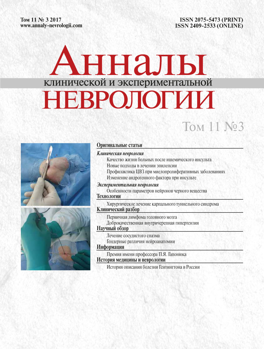
Surgical treatment of the carpal tunnel syndrome using endoscopic and electrophysiological monitoring
- Authors: Vershinin A.V.1, Gushcha A.O.1, Arestov S.O.1, Nizametdinova D.M.1
-
Affiliations:
- Research Center of Neurology
- Issue: Vol 11, No 3 (2017)
- Pages: 41-46
- Section: Technologies
- Submitted: 28.09.2017
- Published: 28.09.2017
- URL: https://annaly-nevrologii.com/journal/pathID/article/view/487
- DOI: https://doi.org/10.17816/ACEN.2017.3.6
- ID: 487
Cite item
Full Text
Abstract
Introduction. Carpal tunnel syndrome (CTS) is a variant of tunnel neuropathy, which develops as a result of compression of the median nerve by a hypertrophic flexor retinaculum. Surgical treatment implies dissection of the flexor retinaculum which leads to fast pain alleviation and termination of neurologic deficit progression.
Objective. To evaluate effectiveness of the new surgical treatment of CTS using endoscopic and electrophysiological monitoring.
Materials and methods. Outcomes of the surgical treatment with the new combined technique were evaluated in a group of 72 patients. To assess effectiveness, VAS, frequency of complications and relapses, length of inpatient hospitalization, and temporary disability were assessed.
Results. We found a significant reduction in VAS pain score from 6 [3; 7] to 2 [1; 3] points within the first day following surgery along with improvement of the surface pain sensitivity from 3 [2; 4] to 2 [2; 3] points. No significant complications of relapses were found (N = 0). The average period of inpatient hospitalization was 16 [12; 24] hours and the temporary incapacity for work was 7 [5; 12] days.
Conclusions. The new surgical approach significantly reduces level of pain syndrome and sensory disturbances, allows to achieve sufficient decompression of the nerve with minimal risks of complications, and reduce duration of hospitalization and temporary disability.
About the authors
Andrey V. Vershinin
Research Center of Neurology
Author for correspondence.
Email: dr.vershinin@gmail.com
Russian Federation, Moscow
Artem O. Gushcha
Research Center of Neurology
Email: dr.vershinin@gmail.com
Russian Federation, Moscow
Sergey O. Arestov
Research Center of Neurology
Email: dr.vershinin@gmail.com
Russian Federation, Moscow
Dinara M. Nizametdinova
Research Center of Neurology
Email: dr.vershinin@gmail.com
Russian Federation, Moscow
References
- Belova N.V., Yusupova D.G., Lagoda D.Yu et al. [Current concept on the diagnosis and treatment of carpal tunnel syndrome]. Russkiy meditsinskiy zhurnal 2015; 23: 1429–1432.
- Marie P., Foix C. Atrophie isolee de l’eminence thenar d’origine nevritique, role du legamente anulaire anterieur du carpe dans la pathogenie de la lesion Revue. Neurology 1913: 21-647.
- Learmoth J.R. The principle of decompression in the treatment of certain deseases of peripheral nerves. Surgical Clinics of North America 2000; 13: 905-933.
- Bertolotti P. Sindromi da intrappolamento dell arto superior. Fondazione Savonese per gli studi sulla mano 1993; 2: 81–121.
- Taleisnik J. The palmarcutaneous branch of the median nerve and the approach to the carpal tunnel. Journal of Bone and Joint Surgery 1973; 55A: 121. PMID: 4758035
- Luchetti R., Amadio P. (eds.) Carpal Tunnel Syndrome. Springer-Verlag Berlin Heidelberg. 2007: 405 p. doi: 10.1007/978-3-540-49008-1
- Crandal R.E., Weeks P.M. Multiple nerve disfunction after carpal tunnel release. Journal Hand Surgery 1988;13A: 584 – 589. PMID: 3418066
- Amadio P.C. The Mayo Clinic and Carpal Tunnel syndrome. Mayo Clinic Proceedings 1992; 67: 42-50. PMID: 1732691
- Lanz U. Anathomical variations of the median nerve in the carpal tunnel. Journal Hand Surgery 1977; 2: 53. PMID: 839054
Supplementary files


