


Complication of COVID-19: Mild Encephalopathy Syndrome with Reversible Splenial Lesion
- Authors: Matveeva T.V.1, Gaifutdinov R.T.1, Kamalova D.S.1, Fasakhova G.A.2
-
Affiliations:
- Kazan State Medical University
- Central City Clinical Hospital No. 18
- Issue: Vol 18, No 4 (2024)
- Pages: 110-116
- Section: Clinical analysis
- Submitted: 28.04.2023
- Accepted: 29.07.2023
- Published: 06.12.2024
- URL: https://annaly-nevrologii.com/pathID/article/view/983
- DOI: https://doi.org/10.17816/ACEN.983
- ID: 983

Cite item

Abstract
A syndrome of mild encephalopathy with reversible splenial lesion (MERS) was described in a post-COVID-19 male patient. The clinical manifestations included neuropsychiatric and visual abnormalities; when focusing separately on an object (one eye closed), the left eye perceived it as normal, but the right eye perceived it as multiple images moving diagonally into the distance. T2, FLAIR, and ADC magnetic resonance imaging (MRI) showed a splenial lesion that resolved rapidly without using corticosteroids. The patient was diagnosed with cerebral polyopia because he saw images arranged in ordered rows after focusing on an object. Differential diagnoses included astigmatism, palinopsia, and polyopic visual hallucinations. Monocular polyopia is explained by anomia associated with the patient's partial split-brain syndrome (the splenial lesion, neuropsychiatric abnormalities); involvement of the pathways from the frontal eye fields to the brainstem structures responsible for initiating extraocular eye movements. The association of neurological complications with prior COVID-19, rapid resolution of symptoms, and MRI lesions without initiating immunosuppressive therapy suggested endotheliopathy as the cause of COVID-19 complications.
Full Text
Introduction
Neurological complications of the novel coronavirus infection (COVID-19) are common, ranging from headache to clinical symptoms and signs of encephalitis, meningitis, encephalomyelitis, acute stroke, Guillain–Barré syndrome, neuropathies, etc. [1–5]. This suggests that their origin is complex and unclear. The possibility of direct cell infection by coronaviruses is under discussion. Angiotensin-converting enzyme 2 and membrane-bound serine protease matriptase-2 are thought to be viral receptors and entry points for some coronaviruses [6, 7]. SARS-CoV-2 enters the brain by the neurogenic route via the axons of olfactory cells and then spreads transsynaptically to various brain structures [8]. The hematogenous spread of the virus is mediated by infected monocytes and macrophages, which contributes to the damage of brain vessel endothelium [9], affecting the function of the neurogliovascular unit (neuron–astrocyte–vessel). This includes an increased permeability of the blood–brain barrier, leading to extravasation of plasma components into the vascular wall and perivascular space, inflammation, loss of autoregulatory function of the brain, smooth muscle involvement, and ultimately occlusion of the vascular lumen at the final stage of the disease [10].
Angiotensin-converting enzyme 2 receptors are found in skeletal muscles, arterial and venous endothelial cells, arterial smooth muscle cells of many organs and the brain [3, 11, 12]. The somatic manifestations and other complications of COVID-19 can be explained by the effects of the viral toxin as well as by inducing effect of the pathogen, leading to a systemic inflammatory response syndrome. A cytokine storm is one of its components. High levels of proinflammatory cytokines (interleukin-1β, -2, and interleukin-2, -4, -10, -18 receptors, interferon-γ, C-reactive protein, tumor necrosis factor-α, granulocyte colony growth factor, etc.) have been reported in severe COVID-19 [2]. However, a retrospective cohort study conducted by M.L. Ciampa et al. in patients hospitalized with COVID-19 did not confirm these data [13]. The difficulty in assessing the role of cytokines in the development of COVID-19 is that cytokines have different functions due to their biological activity. In COVID-19, most conclusions are based on a quantitative assessment of cytokines without considering their functional purpose as substances that activate (stimulate) or suppress (inhibit) the immune response. Considering these factors, the role of cytokines in the development of COVID-19 needs to be clarified.
SARS-CoV-2, like dengue virus, is thought to infect the endothelium directly, resulting in extensive vascular involvement that occurs 3–6 days after disease onset [14, 15]. Endotheliopathy in COVID-19 increases the release of multimeric von Willebrand Factor (VWF) and platelet adhesion, and decreases levels of anticoagulant proteins on the endothelial surface. These processes, together with coagulopathy and infection-induced platelet hyperactivity, trigger blood clot formation and thrombotic microangiopathy [16]. One of the mechanisms that lead to the central nervous system damage in COVID-19 is local hemostasis defects due to an imbalance between high levels of highly active VWF multimers and low levels of ADAMTS13 (a disintegrin and metalloproteinase with thrombospondin motifs 13), which cleaves newly released highly active VWF multimers. In patients with sepsis and a significant imbalance between VWF and ADAMTS13, systemic thrombotic microangiopathy is observed. The imbalance of systemic coagulation factors in COVID-19 patients is mild and may manifest locally. However, it may be one of the reasons for delayed strokes or structural brain lesions [16]. The SARS-CoV-2 induces autoimmune responses by several mechanisms such as molecular mimicry between viral proteins and host antigens, formation of viral superantigens, activation of macrophage and monocyte defense mechanisms, and polyclonal activation of B cells [17].
Viral neuroinvasion can contribute to the exacerbation and progression of acquired, inherited, demyelinating, metabolic, neurodegenerative, and neuromuscular diseases [2, 18–20]. It is still unclear how the underlying mechanism for a particular form of the disease can be identified.
A case report
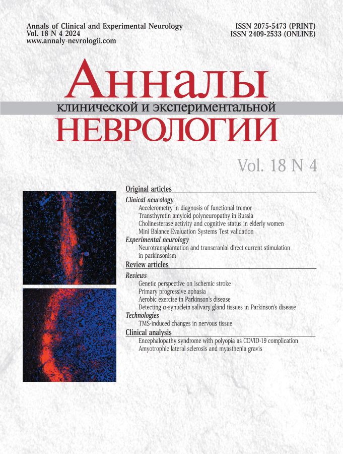
Patient A., 31 years old, was admitted with complaints of visual impairment. When looking with both eyes, he saw an object as “a set of identical objects arranged in a row” (Figure 1). When the patient covered his left eye, the right eye's perception remained distorted. With the right eye closed, the left eye's perception was normal. The same visual disturbances occurred when refocusing on a new object. The object and background were clearly seen. Duration of visual impairment was approximately 1 minute.

Fig. 1. This is how patient A. saw when he fixes his gaze on something.
The patient noted gait instability, impaired memory for recent events (did not remember if he took the drug the day before or at the day of examination). The patient felt constantly anxious, detached from his surroundings, fearful of the future. It seemed as if “everything was happening not to” him. This experience was accompanied by general weakness, sweating, and a blood pressure increase to 160/90 mm Hg.
Visual impairment occurred on day 21 after moderate COVID-19. After being discharged from COVID-19 department, he could not return to work due to his inability to concentrate and perform routine tasks. He became more anxious. Prior to this case, he rarely had acute respiratory viral infections.
The neurological examination on admission to the Neurology department did not reveal any visual fields deficit in confrontation test. No sensory or motor disorders were observed. Deep tendon reflexes were active and symmetrical. Abdominal reflexes were absent, plantar reflexes were reduced. No abnormal signs were detected. Coordination tests revealed slight bilateral dysmetria. Bilateral dysdiadochokinesis was inconclusive. On Romberg test, the patient swayed slightly. Gait was unsteady, tandem walking was normal. A Montreal Cognitive Assessment Scale score was 27. The patient was unable to accurately redraw the cube and reproduce two words by heart (but was able to do so when prompted by category). In a ten-word test, the patient could recall only 4 of 10 words after 1 repetition. After the subsequent repetitions, he could reproduce 7, then 9, 9, and all 10 words. The volume of a correction task was 583 characters. A concentration score was 4.85. High attention span, decreased work capacity, and significant emotional instability were observed.
Laboratory tests showed ALT of 53 U/L (reference: < 37 U/L), AST of 29 U/L (reference: < 29 U/L), ferritin of 272 ng/mL (reference: 2–250 ng/mL). Coagulation test was unchanged. Other parameters were normal. The ophthalmologist's conclusion was simple astigmatism of the left eye, bilateral retinal angiopathy.
Magnetic resonance imaging (MRI) of the brain showed a hydrophilic splenial lesion measuring 12 × 18 × 15 mm without perifocal changes or contrast enhancement, consistent with cytotoxic lesions of the corpus callosum (CLOCCs; Figures 2–4).

Fig. 2. Axial brain FLAIR MRI of Patient A.
A: day 1 of hospitalization; a single symmetric hyperintense splenial lesion with relatively smooth and clear contours; B: day 13 of hospitalization; lesion almost completely resolved.

Fig. 3. ADC Map MRI of Patient A.
A: day 1 of hospitalization, low signal intensity in the splenium projection; B: day 13 of hospitalization; average signal intensity in the splenium projection.

Fig. 4. Sagittal brain FLAIR MRI of рatient A.
A: day 1 of hospitalization; hyperintense splenial lesion; B: day 13 of hospitalization, decreased intensity of the lesion.
The patient received metabolic therapy. On day 4 of hospitalization, visual disturbances resolved. The patient was discharged in satisfactory condition.
Discussion
In our case, the visual impairment could not be explained by the ophthalmologist's diagnosis of astigmatism, with its main symptoms being blurred and double vision. The patient clearly saw multiple images of objects arranged in a row.
This visual impairment was classified as cerebral polyopia because of its dependence on gaze fixation and its specific character. Polyopia is seeing two or more images arranged in ordered rows, columns, or diagonals after fixation on an object [21].
Cerebral polyopia differs from ocular polyopia, which is associated with the formation of areas in the optical media of the eye (cornea, lens) that refract light rays unevenly, resulting in the projection of multiple object images onto the retina. In our case, one of the objects could be perceived clearly, while the other could appear blurred. The visual defect was not associated with fixation, persisted when one eye was closed, and did not disappear when the patient was attempting to look at an object using a needle-hole occluder.
The patient's visual impairment also differed from palinopsia. Palinopsia is a visual perseveration. Patients continue to perceive or see the image again for a brief time even after the visual stimulus stops. The virtual image is perceived as real in the environment and does not disappear when the eyes are closed. Illusion occurs on the side of the defective visual field. Palinopsia occurs when the process is located in the temporo-occipital, parietal-occipital regions of the brain and, less commonly, in the posterior regions of the left hemisphere [22].
Cerebral polyopia is observed in cases of damage to the occipital or parieto-occipital regions of the brain [23] or to the parietal region [24]. The neurophysiological mechanism of polyopia is associated with increased excitability of neurons in the visual cortex [25]; with recoding of visual receptive fields in the primary visual cortex with bilateral lesions in the occipital lobe [26]; with disruption of connections between the posterior parietal cortex, where visual-spatial analysis is performed, and the cortical gaze center, subcortical structures, and the stem gaze center [24]. Due to the lack of neuroimaging data confirming brainstem and hemisphere damage, preservation of visual fields, and monocular character of the defect, visual impairment is unlikely to be explained by the above mechanisms.
The dependence of the visual impairment on fixation and the monocular polyopia associated with an existing object excludes the possibility of visual polyopic hallucinations with the virtual image seen [22].
Foveal fixation and stereopsis (the sense of spatial expansion and relief of real objects) are performed by each eye separately. Perception of an object by a person with normal binocular vision requires precise alignment of the visual axis to re-fixate the object in each eye in the same dimension, which is achieved by extraocular (vergent) movements of the eyeballs (saccades) [27]. Visual information enters the primary visual field, the striate cortex or visual area V1, whose axons form the dorsal and ventral visual pathways. The dorsal pathway provides the answer to the question “Where?”, terminates in neurons located in the posterior parietal region, and is associated with the spatial orientation of objects. The ventral pathway provides the answer to the question “What?”, is associated with the identification of objects, and is adapted to the structures of the extrastriate visual cortex (V2, V3, V4, V5 fields). From the parietal region, the information reaches the frontal eye fields, which generate a targeted motor effect of the eyeballs in the form of saccades. At the same time, the role of the frontal eye fields is reduced to selecting the appropriate saccadic amplitude and transmitting the information through a number of intermediate structures, including the reticular and paramedian structures of the brainstem, to the muscles that provide extraocular eye movements [24, 27]. In our case, the patient clearly saw and recognized the object, and this suggested the spared ventral visual pathway. This allowed concluding that the patient's image perception was associated with a defect in foveal fixation and stereopsis of the object. Given the location of the process, it can be assumed that pathways from the frontal fields of the eye to the brainstem were affected. However, we cannot explain monopolyopia by a disruption of the considered connections.
In our case, monopolyopia was accompanied with a splenial lesion, which suggested the possibility of a partially split-brain syndrome associated with damage to the callous structures. This was confirmed by the experiment of Gazzaniga, 1999 [28]. When an object was presented to the left hemisphere, a split-brain patient gave the correct answer with an emotional response. When the same object was presented to the right hemisphere, the patient responded that she saw nothing. In cases of combined damage to the visual pathways and centers, partial transection of the posterior corpus callosum resulted in visual anomia [28]. This is the most likely defect in our patient.
Lesions in the isthmus/splenium and corpus callosum are associated with confusion, altered mental status, hallucinations, psychosis, mutism, and cognitive impairment [29–31]. This location may explain impaired anterograde memory, anxiety, fear, emotional instability, impaired auditory short-term memory (with less words retained and reproduced for the first sequence of the Luria test); depersonalization and derealization.
In our case, instability of walking cannot be explained by cerebellar symptoms. It does not correlate with inconclusive cerebellar symptoms, normal tandem walking, and unsteady but not ataxic gait. Instability is one of the corpus callosum lesion symptoms, with some patients unable to move [28].
There are acute and chronic variants of the split-brain or disconnection syndrome. Acute symptoms of disconnection may develop gradually or rapidly, within 4–7 days, and may partially or completely resolve [32–34], as in our patient.
Prior COVID-19, as well as the corresponding described symptoms suggested the diagnosis of mild encephalopathy with reversible splenial lesion (MERS) associated with COVID-19; partially split-up brain syndrome in the form of transient mononuclear cerebral polyopia, visual anomia, and mild neuropsychiatric disorders.
Based on the above-mentioned mechanisms of nervous system damage in SARS-CoV-2 infection, the presence of CLOCCs, the absence of toxic infectious manifestations or hemostasis disorders on admission to the Neurology department, rapid regression of visual impairment without using steroids (the patient received only metabolic therapy) suggest the vascular nature of the disease and associates the resulting condition with local endotheliopathy, which is possible in COVID-19 [16, 35] and may be the cause of CLOCCs [29].
MERS is a new disease entity with limited experience in diagnosing and understanding clinical manifestations.
We detected and confirmed monocular cerebral polyopia and visual anomaly in the patient with the splenial lesion. According to our hypothesis, these syndromes are thought to develop due to the splenial lesion and partial damage to the callous pathways and descending visual connections that pass near the corpus callosum. These syndromes in patients with lesions of the corpus callosum have not been previously described, so this information may contribute to the understanding of the function of the posterior corpus callosum.
There are almost no reports of MERS in COVID-19, so this report may be interesting for clinicians and brain researchers.
About the authors
Tatiana V. Matveeva
Kazan State Medical University
Email: gaifutdinov69@mail.ru
ORCID iD: 0000-0002-1889-0094
Dr. Sci. (Med.), Prof., Department of neurology, neurosurgery and medical genetics
Russian Federation, KazanRustem T. Gaifutdinov
Kazan State Medical University
Author for correspondence.
Email: gaifutdinov69@mail.ru
ORCID iD: 0000-0001-5591-7148
Cand. Sci. (Med.), Associate Professor, Department of neurology, neurosurgery and medical genetics
Russian Federation, KazanDinara S. Kamalova
Kazan State Medical University
Email: gaifutdinov69@mail.ru
ORCID iD: 0000-0002-3123-9546
neurologist, Central City Clinical Hospital No. 18
Russian Federation, KazanGulnaz A. Fasakhova
Central City Clinical Hospital No. 18
Email: gaifutdinov69@mail.ru
ORCID iD: 0009-0004-4843-6767
Head, Neurology department
Russian Federation, KazanReferences
- Белопасов В.В., Яшу Я., Самойлова Е.М., Баклаушев В.П. Поражение нервной системы при СOVID-19. Клиническая практика. 2020;11(2):60–80. Belopasov V.V., Yashu Ya.A., Samojlova E.M., Baklaushev V.P. Damage to the nervous system in COVID-19. Clinical practice. 2020;11(2):60–80. doi: 10.17816/clinpract34851
- Громова О.А., Торшин И.Ю., Семенов В.А. и др. О прямых и косвенных неврологических проявлениях CОVID-19. Журнал невропатологии и психиатрии им. С.С. Корсакова. 2020;120(11):11–21. Gromova O.A., Torshin I.Yu., Semenov V.A. et al. On the direct and indirect neurological manifestations of COVID-19. Journal of Neuropathology and Psychiatry named after S.S. Korsakov. 2020;120(11):11–21. doi: 10.17116/jnevro 202012011111
- Mao L., Jin H., Wang M. et al. Neurologic manifestations of hospitalized patients with coronavirus disease 2019 in Wuhan, China. JAMA Neurol. 2020;77(6):683–690. doi: 10.1001/jamaneurol.2020.1127
- Stafstrom C.E., Jantzie L.L. COVID-19: neurological considerations in neonates and children. Children (Basel). 2020;7(9):133. doi: 10.3390/children7090133
- Suri V., Pandy S., Sing J., Jena F. Acute onset chronic inflammatony demyelinating polyneupathy after COVID-19 infection and subsequent ChAdOx1 nCoV-19 vaccination. Case Rep. 2021;14:e245816. doi: 10.1136/dcr-2021-245816
- Ennaji M.M. Emerging and reemerging viral pathogens. Vol. 1: Fundamental and basic virology aspects of human, animal and plant pathogens. London; 2020.
- Bandala C., Cortes-Altamirano J.L., Reyes-Long S. et al. Putative mechanism of neurological damage in COVID-19 infection. Acta Neurobiol. Exp. (Wars). 2021;81(1):69–79. doi: 10.21307/ane-2021-008
- Gandhi S., Srivastava A.K., Ray U., Tripathi P.P. Is the collapse of the respiratory center in the brain responsible for respiratory breakdown in COVID-19 patients? ACS Chem. Neurosci. 2020;11(10):1379–1381. doi: 10.1021/acschemneuro.0c00217
- Zhou Z., Kang H., Li S., Zhao X. Understanding the neurotropic characteristics of SARS-CoV-2: from neurological manifestations of COVID-19 to potential neurotropic mechanisms. J. Neurol. 2020; 267(8):2179–2184. DOI: 10/1007/s00415-020-09929-7
- Wardlaw J.M., Smith C., Dichgans M. Mechanisms of sporadic cerebral small vessel disease: insights from neuroimaging. Lancet Neurol. 2013;12(5):483–497. doi: 10.1016/S1474-4422(13)70060-7
- Baig A.M., Khaleeq A., Ali U., Syeda H. Evidence of the COVID-19 virus targeting the CNS: tissue distribution, host-virus interaction, and proposed neurotropic mechanisms. ACS Chem. Neurosci. 2020;11(7):995–998. doi: 10.1021/acschemneuro.0c00122
- Hamming I., Timens W., Bulthuis M.L. et al. Tissue distribution of ACE2 protein, the functional receptor for SARS coronavirus. A first step in understanding SARS pathogenesis. J. Pathol. 2004;203(2):631–637. doi: 10.1002/path.1570
- Ciampa M.L., O’Hara T.A., Joel C.L. et al. Absence of “cytokine storm” in hospitalized COVID-19 patients: a retrospective cohort study. Infect. Dis. Rep. 2021;13(2):377–387. doi: 10.3390/idr13020036
- Prasad M., Leon M., Lerman L.O., Lerman A. Viral endothelial dysfunction: a unifying mechanism for COVID-19. Mayo Clin Proc. 2021;96(12):3099–3108. doi: 10.1016/j.mayocp.2021.06.027
- Vervaeke P., Vermeire K., Liekens S. Endothelial dysfunction in dengue virus pathology. Rev. Med. Virol. 2015;25(1):50–67. doi: 10.1002/rmv.1818
- Portier I., Campbell R.A., Denorme F. Mechanisms of immunothrombosis in COVID-19. Curr. Opin. Hematol. 2021;28(6):445–453. doi: 10.1097/MOH.0000000000000666
- Shabani Z. Demyelination as a result of an immune response in patients with COVD-19. Acta Neurol. Belg. 2021;121(4):859–866. doi: 10.1007/s13760-021-01691-5
- Копишинская С.В., Жаринова Н.О., Величко И.А. и др. Основные принципы ведения неврологических пациентов в период пандемии COVID-19. Нервно-мышечные болезни. 2020;10(1):31–42. Kopishinskaya S.V., Zharinova N.O., Velichko I.A. et al. Basic principles of neurological patient management during the COVID-19 pandemic. Nervnomyshechnye bolezni. 2020;10(1):31–42. DOI: 10.17650 /2222-8721-2020-10-1-31-42
- International MG/COVID-19 Working Group, Jacob S., Muppidi S. et al. Guidance for the management of myasthenia gravis (MG) and Lambert–Eaton myasthenic syndrome (LEMS) during the COVID-19 pandemic. J. Neurol. Sci. 2020;412:116803. doi: 10.1016/j.jns.2020.116803
- Rajabally Y.A., Goedee H.S., Attarian S., Hartung H.P. Management challenges for chronic dysimmune neuropathies during the COVID-19 pandemic. Muscle Nerve. 2020;62(1):34–40. doi: 10.1002/mus.26896
- Jones M.R., Waggoner R., Hoyt W.F. Cerebral polyopia with extrastriate quadrantanopia: report of a case with magnetic resonance documentation of V2/V3 cortical infarction. J. Neuroоphthalmol. 1999;19(1):1–6. doi: 10.1097/00041327-199903000-00001
- Brazis Pol U., Med'yu D.K., Billert H. Topical diagnosis in clinical neurology. Philadelphia; 2001.
- Isherwood S., Jewsbury H., Nitkunan A., Ali N. An unusual case of cerebral polyopia. Can. J. Ophthalmol. 2017;52(3):e102–e104. doi: 10.1016/j.jcjo.2016.10.016
- Kesserwani H. A novel case of cerebral diplopia secondary to a posterior parietal cortex ischemic infarct: proposal of a mechanism of generation of polyopia due to convergence insufficiency. Cureus. 2021;13(1):e12962. doi: 10.7759/cureus.12962
- Gersztenkorn D., Lee A.G. Palinopsia revamped: a systematic review of the literature. Surv. Ophthalmol. 2015;60(1):1–35. doi: 10.1016/j.survophthal.2014.06.003
- Cornbrath W.T., Butter C.M., Barnes L.L. et al. Spatial characteristics of cerebral poliopia: a case study. Vision Res. 1998;38(24):3965–3978. doi: 10.1016/s0042-6989(97)00431-8
- Searle A., Rowe F.J. Vergence neural pathways: a systematic narrative literature review. Neuroophthalmology. 2016;40(5):209–218. doi: 10.1080/01658107.2016.1217028
- Козявина М.С. Нейропсихологический анализ патологии мозолистого тела. М.; 2012. Kozyavina M.S. Neuropsychological analysis of cerebellar body pathology. Moscow; 2012. (In Russ.)
- Перов Р.И., Хакимова А.Р., Попова Н.А. Cиндром умеренной энцефалопатии с обратимым поражением валика мозолистого тела: обзор литературы и собственное наблюдение в неотложной неврологической клинике. Вестник современной клинической медицины. 2018;11(5):109–114. Perov R.I., Hakimova A.R., Popova N.A. The syndrome of moderate encephalopathy with reversible lesions of the corpus callosum: a review of the literature and our own observation in an emergency neurological clinic. Bulletin of modern clinical medicine. 2018;11(5):109–114. doi: 10.20969/VSKM.2018.11(5).109-114
- Doherty M.J., Jayadev S., Watson N.F. et al. Clinical implications of splenium magnetic resonance imaging signal changes. Arch. Neurol. 2005;62(3):433–437. doi: 10.1001/archneur.62.3.433
- Park M.K., Hwang S.H., Jung S. et al. Splenial lesions of the corpus callosum: disease spectrum and MRI findings. Korean J. Radiol. 2017;18(4):710–721. doi: 10.3348/kjr.2017.18.4.710
- Park M.K., Hwang S.H., Jung S. et al. Lesions in the splenium of the corpus callosum: clinical and radiological implications. Neurol. Asia. 2014;19(1):79–88.
- Jea A., Vachhrajani S., Widjaja E. et al. Corpus callosotomy in children and the disconnection syndromes: a review. Child’s Nerv. Syst. 2008;24(6):685–692. doi: 10.1007/s00381-008-0626-4
- Uda T., Kunihiro N., Umaba R. et al. Surgical aspects of corpus callosotomy. Brain Sci. 2021;11(12):1608. doi: 10.3390/brainsci11121608
- Pristas N., Rosenberg N., Pindrik J. et al. An observational report of swallowing outcomes following corpus callosotomy. Epilepsy Behav. 2021;123:108271. doi: 10.1016/j.yebeh.2021.108271
Supplementary files
