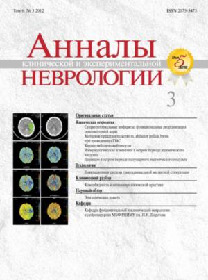
Motor recovery after ischemic stroke is associated with the neural networks reorganization. fMRI studies of these networks in patients with mild motor deficit showed that activation pattern can be used for the prognosis of functional recovery. However, characteristics and clinical relevance of activation patterns in patients with severe to moderate motor deficit, its effective functioning in patients with different severity of primary sensorimotor system components (corticospinal tract [CST] and primary sensorimotor [SM] cortex) injury were not investigated. Twentyfive chronic hemispheric ischemic stroke patients were studied (13 males, 12 females; median age 38.0±5.9 years). Depending on the severity of hand motor impairment and functional outcome the patients were divided into 3 groups. All patients underwent 1.5 T fMRI (Siemens Avanto) with passive block paradigm of paretic index finger movement (1 Hz frequency). Statistic maps of group activation showed significant differences in groups with different functional outcome: the more severe was motor deficit, the less SM activation volume size in injured hemisphere was seen, and activation cluster center moved towards non-primary motor cortex. The association between the activation volume of SM and structural integrity of CST, assessed by fractional anisotropy index was revealed. Statistic maps of individual activation showed SM activation in injured hemisphere in 38% patients with unfavourable (severe paresis, plegia) and moderate recovery with different physiologic lateralization, that is typical for the group with good recovery (mild and moderate paresis). These data do not allow us to consider the activation pattern as a marker of motor recovery and prognostic factor in patients with severe and moderate motor deficit. Our results showed that sensorimotor networks formation and functioning differ depending on the CST sparing, and its effective work is possible in certain degree CST integrity.
Vol 6, No 3 (2012)
- Year: 2012
- Published: 10.09.2012
- Articles: 8
- URL: https://annaly-nevrologii.com/journal/pathID/issue/view/24
Full Issue
Original articles
 4-13
4-13


Сortical mapping of m. abductor polliсis brevis motor area in healthy volunteers using navigation transcranial magnetic stimulation
Abstract
Transcranial magnetic stimulation (TMS) is method of brain neurons stimulation by alternating magnetic field and measuring motor-evoked potentials (MEPs) with electromyography monitoring. Recently, some new technologies were introduced, based on classical TMS, which allowed noninvasive brain mapping of cortical motor areas. Navigation transcranial magnetic stimulation (nTMS, NBS eXimia Nexstim) is one of them. In this article we describe our experience of m. abductor pollicis brevis motor area mapping in 29 healthy volunteers. Basic parameters of TMS were measured: MEP amplitude (408.49±216.36 uV), latency (22.38±0.97 ms), passive motor threshold (40.08±4.68% for the left hemisphere, 48.71±9.16% for the right hemisphere). Maps of cortical areas were builded.
 14-17
14-17


Cardioembolic stroke: characteristics of cerebral, systemic and intracardial hemodinamics and their interrelations with lesion lateralization
Abstract
In this clinical-instrumental study abnormalities of blood pressure circadian changes were described. High incidence of ventricle arrhythmias and episodes of latent AV-nodal conduction was revealed. Features of structural-functional heart condition and its interrelation with myocardial electrical instability were studied. Incidence of atherosclerotic lesion, stenosis degree, occurrence of hypoechogenic plaques were analyzed. Close relationship between myocardial electrical instability, structural-functional heart condition and cerebral blood flow were assessed. These interrelations indicated the connection between heart and cerebral lesion in patients with hypertension and atrial fibrillation. The severity of these lesions and the degree of cardiocerebral interrelations increase in ischemic stroke, especially in right-sided ischemic lesions.
 18-24
18-24


Immunological changes in acute ischemic stroke
Abstract
The pattern of changes of cellular and humoral immunity were studied in 41 patients with acute ischemic stroke with assessment on 1st–3rd, 7th, 21st days. Control group included 31 patients with arterial hypertension. At 1st–3rd days of stroke lymphopenia-deficient CD3+, CD4+ and CD8+ cells was detected, in 33% of patients the reduction of IgG was found. On Day 7 and Day 21 some positive changes were noted, however, in large proportion of patients symptoms of immunodeficiency with reduction in the number of CD8+ and RI index remained. These findings suggest that the immune status of patients in acute stroke is consistent with response on acute stress. The mechanisms of immune system involvement in pathogenesis of acute stroke are discussed.
 25-30
25-30


Citicoline (Ceraxon) in acute stroke: assessment of clinical efficacy and effects on cerebral perfusion
Abstract
Novel neuroimaging techniques provide quantitative assessment of cerebral perfusion in acute stroke and reveal the heterogeneity of ischemic zone. Neuroprotective agents play major role in the treatment of acute stroke as they are intended to restore functioning of potentially viable tissue. This prospective open-label study included 50 patients (mean age 60.9 years) with acute hemispheric stroke within the first 24 hours of symptoms onset. Patients were divided into 2 arms (25 patients in each arm) to receive standard of care (control arm) or standard of care plus citicoline (Ceraxon) 1 g b.i.d. as I.V. injection for 10 days. Clinical symptoms were assessed with NIHSS; neuroimaging included DWI to confirm ischemic lesion and perfusion CT to assess cerebral perfusion. Patients in both arms demonstrated significant clinical improvement on Day 10 with no significant difference between treatment arms (mean NIHSS score was 9.4 in control arm and 8.4 in Ceraxon arm, p=0.87). Perfusion CT on admission showed perfusion deficit in all patients. Mismatch regions on perfusion CT compared to DWI indicating potentially viable tissue (“penumbra”) were found in 75% of patients in control arm and in 69% of patients in Ceraxon arm. No difference between perfusion parameters in the “core” vs. “penumbra” on initial imaging was shown. On Day 10 there were no changes of cerebral perfusion values in the “core” regions, while in “penumbra” in Ceraxon arm CBF increased significantly CBF (p=0.013) with no significant differences vs. intact hemisphere, that is consistent with cerebral perfusion improvement. Thus, treatment with Ceraxon in the first 10 days of acute stroke may result in improvement of cerebral perfusion in the potentially viable tissue.
 31-36
31-36


Reviews
Episodic memory: neurological and neuromediator mechanisms
Abstract
Significant advances in understanding of neurological and neuromediator mechanisms of memory along with the causes of memory decline in aging were achieved recently. Functioning of episodic memory system needs considerable energetic and material (neuromediator, protein) resources, and appears to be highly consuming for the body. Therefore, physiological mechanisms inhibiting episodic memory system exist in order to distribute resources for other brain functions. Primary engram is recorded by hippocampal structures, which maintain reciprocal connectivity with neocortex zones involved in synchronized activity. Cholinergic innervation of hippocampus and primary zones of neocortex stabilizes process of primary engram formation with consequent transformation of synapses in hippocampus structures. At the next stage hippocampus transfers the engram to neocortex, where the information is processed by associating with previous knowledge and is fixed by slowly developing synaptogenesis and axonal growth. Noradrenergic innervation is an essential part of neuroplastic processes. Possible causes of memory decline in aging include powerful influences of mature prefrontal cortex, inhibiting the hippocampal activity; insufficient stimulation of hippocampal memory system by novel events with consequent decline of neurogenesis and neuroplasticity; disturbances in mechanisms of phasic release and
clearance of neuromediators.
 53-60
53-60


Technologies
New step to a personalized medicine. Navigation transcranial magnetic stimulation (NBS eXimia Nexstim)
Abstract
Navigation transcranial magnetic stimulation of the brain (nTMS) NBS eXimia Nexstim – new method based on the stimulation of neurons by an alternating magnetic field and recording responses to stimulation with electromyography. Precise guidance of the stimulus to a particular area of the cerebral cortex the patient, repeat the stimulus, somatotopical mapping of motor areas are features of this method. These characteristics are achieved by a special navigation system that compares patient’s head with the anatomical MRI image and the location of stimulating magnetic coil. This review presents the current possibilities of the navigation TMS method in the diagnosis and treatment of neurological diseases; our own local experience with the system.
 37-46
37-46


Clinical analysis
Comorbidities in cerebrovascular diseases
Abstract
Cerebrovascular diseases are the major cause of morbidity and mortality among adults. The extensive development of various diagnostic methods and their introduction into routine practice increased the detection of previously underappreciated and unknown neurological diseases. Comorbidity, i.e. the joining of various other disorders of nervous system (neurodegenerative or demyelinating) to cerebrovascular disease, often leads to a layering of additional symptoms which contribute to a significant decline of the quality of life and increase the costs of treatment. In this clinical analysis case histories of patients with combined disorders of the nervous system are reviewed. Approaches to the diagnostic strategy and the need for personalized treatment based on the identified comorbidity are discussed.
 47-52
47-52


