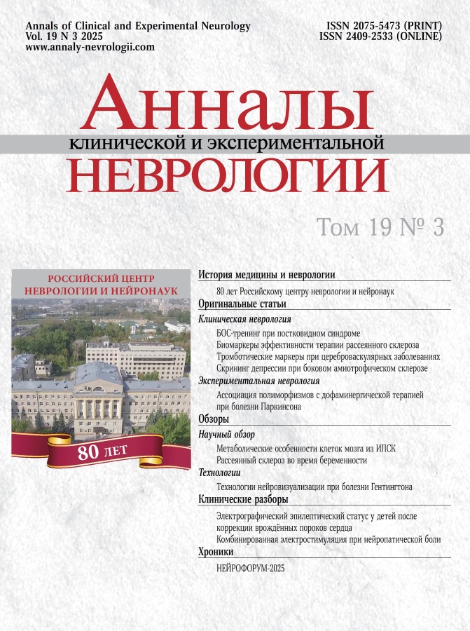
Annals of Clinical and Experimental Neurology
Peer-review quarterly medical journal.
Editor-in-Chief
- Prof. Michael Piradov
ORCID: 0000-0002-6338-0392
Founders
Publisher
About
The journal “Annals of Clinical and Experimental Neurology” is a peer-review medical journal, which provides articles for practicing neurologists, neurosurgeons, cardiologists, critical care and neurorehabilitation professionals, neuropsychologists, neuroradiologists, clinical neurophysiologists, as well as neuroscience professionals.
The journal publishes original articles, scientific reviews on all problems of central and peripheral nervous system diseases, fundamental neurosciences, and also on problems adjacent to other medical specialties. In addition, the journal accepts technology reviews in neurology and neurosciences, clinical studies and essays on the history of neuroscience and neuroscience.
The journal’s unique mission is to cover current problems and modern achievements in the field of neurology, neurosurgery, critical care, neurorehabilitation, neuroimaging, cardioneurology, clinical neurophysiology, fundamental neuroscience as well as to contribute to the formation of new promising research and training of highly qualified personnel in these areas.
Journal’s main tasks are:
- Reflection of the results of scientific research in the most significant areas of neurology and related neuroscience
- Regular informing of the medical community about the latest achievements and prospects for the development of domestic and foreign medical science in the field of neurology
- Promoting the widespread introduction into practice of the latest achievements of neuroscience
- Creation of a platform for an exchange of views on the most significant problems of clinical and fundamental neuroscience, professional development and knowledge level of specialists
APC, Publication & Distribution
- Quarterly issues (4 times a year)
- Platinum Open Access (no APC)
- Creative Commons Attribution 4.0 International (CC BY 4.0) License.
Indexation
- Scopus
- Russian Science Citation Index (on WoS)
- CrossRef
- DOAJ (Directory of Open Access Journals)
- Google Scholar
- Ulrich’s International Periodicals Directory
Current Issue
Vol 19, No 3 (2025)
- Year: 2025
- Published: 10.10.2025
- Articles: 12
- URL: https://annaly-nevrologii.com/pathID/issue/view/87
Historical articles
On the 80th anniversary of the Russian Center for Neurology and Neuroscience
 6-13
6-13
Original articles
Biofeedback training in rehabilitation of patients with neurological disorders in post-COVID syndrome: a randomized controlled trial
Abstract
Introduction. The high prevalence of post-COVID syndrome (PCS), which frequently manifests with emotional disturbances, cognitive impairment, and asthenia, necessitates effective rehabilitation methods. One potential approach is electroencephalography (EEG)-based biofeedback (BFB) therapy, though its use in PCS management has been explored in only a few studies to date.
The study aimed to evaluate the effects of EEG α-rhythm BFB training on emotional state and cognitive function recovery, and reduction of astheniа symptoms in PCS patients.
Materials and methods. Patients diagnosed with U09. Post-COVID-19 condition were randomly assigned to two groups of 10 participants each. The main group underwent 12–15 sessions of EEG α-rhythm BFB training using the NeuroPlay-6C headset with the Neurocorrection of COVID-19 Psychoemotional Consequences protocol, while the control group received identical training without biofeedback. Assessments performed before and after the intervention included: emotional state evaluation (State-Trait Anxiety Inventory [STAI], Short Health Anxiety Inventory [SHAI], Beck Depression Inventory [BDI], Psychological Stress Measure [PSM-25]), cognitive function assessment (Addenbrooke’s Cognitive Examination III [ACE-III], Schulte tables, Stroop test, Tower of London test, N-back test, 10-word memory test), assessment of asthenia (Multidimensional Fatigue Inventory [MFI]), and sleep quality evaluation (Insomnia Severity Index [ISI]).
Results. In both groups, the training resulted in a significant reduction of personal anxiety, psychological stress, depression, and asthenia. The main group additionally demonstrated decreased health-related anxiety and improved information retention parameters. Intergroup comparison revealed more pronounced dynamics in the main group: greater reduction of general fatigue manifestations, increased immediate word recall volume, and improved retention of verbal information in working memory. The proportion of patients transitioning to milder symptom severity levels on individual scales was comparable between both groups.
Conclusion. EEG α-rhythm biofeedback training can be implemented at the outpatient rehabilitation stage for PCS patients.
 14-26
14-26
Predicting the efficacy of anti-B-cell therapy in patients with multiple sclerosis
Abstract
Introduction. Prognostic markers can be used to evaluate a response to anti-B-cell therapy in patients with multiple sclerosis (MS).
Aim. The study aimed to evaluate the characteristics of peripheral blood (PB) lymphocytes and monocytes in patients with aggressive MS during the first 6 months of anti-B-cell therapy.
Materials and methods. Twenty-nine patients with aggressive MS were treated with a humanized anti-CD20 monoclonal antibody (anti-CD20 mAb). A panel of MAbs to differentiation antigens of PB lymphocytes was used to assess the parameters of cellular immunity using six-color flow cytometry. The reference values were based on the similar parameters of ten apparently healthy volunteers.
Results. At month 6, the initial course of anti-CD20 therapy resulted in low recovery of the PB sub-populations of B cells in 85% of patients. Significant decreases were reported in the absolute counts of T cells, T helper cells, Natural Killer (NK) cells, and relative percentage of natural killer T (NKT) cells. The study also showed low levels of activated T cells and significantly decreased percentage of memory B cells (CD27+) and B cells expressing costimulatory and activation molecules (CD40+, CD38+, and CD25+, respectively). A significant decrease in the mean fluorescence intensity of HLA-DR was observed on PB monocytes compared to normal values and those in patients receiving other disease-modifying therapies. Anti-CD20 therapy may indirectly suppress their antigen-presenting ability. Other immunological criteria for prediction of MS progression and magnetic resonance imaging activity during the first year of anti-B-cell therapy may include the following changes from baseline: increased percentages of CD3+, CD3+HLA-DR+, CD25+CD3+, and CD95+CD3+ cells; significant expression of the CD40 molecule and B-cell activation markers CD38 and CD25, and decreased expression of CD95.
Conclusion. Further research on changes in cellular immunity parameters during anti-CD20 therapy could allow for early adjustment of MS treatment to stabilize the patient’s condition.
 27-36
27-36
Early markers of thrombotic hazard in cerebrovascular diseases
Abstract
Introduction. Cerebrovascular disease (CVD) is a heterogeneous group of difficult-to-diagnose conditions in which hemorheological and hemostatic disorders significantly impact the risk of ischemic stroke (IS), as well as the prognosis and response to reperfusion therapy and preventive treatment. Laboratory thrombotic hazard markers, such as the thrombin-antithrombin III (TAT) complex, the plasmin-α2-antiplasmin (PAP) complex, thrombomodulin (TM), and the tissue plasminogen activator (tPA)/plasminogen activator inhibitor-1 (PAI-1) activity ratio, have not been adequately evaluated as predictors of different IS subtypes. Their potential role in acute IS has also not been determined.
Aim. The study aimed to evaluate the diagnostic and predictive value of primary thrombotic hazard markers in patients with CVD.
Materials and methods. The retrospective study included 91 patients with acute IS (45% of men; median age: 62 years). At admission, primary clinical parameters were assessed, including a National Institutes of Health Stroke Scale (NIHSS) score. Laboratory parameters and thrombotic hazard markers were also measured using an enzyme-linked immunosorbent assay. Three IS subtypes included large artery atherosclerosis (LAA)-related IS (n = 32), lacunar IS (n = 27), and hemorheological (small artery occlusion-related) IS (n = 32). The clinical outcomes were evaluated at day 10 using the NIHSS scale. A comparison group included patients with chronic CVD (n = 29; 34% men; median age: 55 years).
Results. The plasma levels of almost all study biomarkers differed significantly between patients with IS and chronic CVD, as well as between patients with different IS subtypes. Four of six markers (PAI-1, PAP, TAT, t-PA/PAI-1) were significantly associated with IS development, with TAT showing the strongest association (odds ratio: 4.78; 95% confidence interval: 2.70, 9.68). Linear regression models were used to evaluate the predictive value of thrombotic hazard biomarkers for IS outcomes, and TAT showed the most significant association in this case (p < 0.001). An analysis of the differential value of study biomarkers for different IS subtypes showed that PAI-1 was the most sensitive (0.969) marker for LAA-related IS, while t-PA/PAI-1 (0.99) and TAT (0.889) demonstrated high predictive value for lacunar IS.
Conclusion. Thrombotic hazard markers are a promising laboratory tool for evaluating IS risk and predicting functional outcomes and response to reperfusion therapy in patients with IS.
 37-48
37-48
Depression in amyotrophic lateral sclerosis: screening using attention bias assessment
Abstract
Introduction. Depression is highly prevalent in amyotrophic lateral sclerosis (ALS) [9]. Its detection can be challenging, particularly in advanced disease when many patients develop bulbar dysfunction and upper limb muscle weakness. This necessitates objective methods for diagnosing affective disorders in ALS.
The study aimed to evaluate the potential utility of eye-tracking technology for detecting depression in ALS patients using an attention bias assessment.
Materials and methods. The study enrolled ALS patients meeting Gold Coast criteria. Depressive symptoms were assessed using the Hospital Anxiety and Depression Scale (HADS). During eye-tracking sessions, patients viewed a screen displaying pairs of faces — one emotional (sad or happy) and one neutral.
Results. Data from 33 participants were analyzed. Comparative analysis of mean gaze fixation duration showed that ALS patients with depressive symptoms had significantly longer fixation times on sad faces compared to non-depressed patients. HADS depression scores correlated with mean fixation duration on sad (r = 0.421; p = 0.018) and neutral (r = 0.36; p = 0.047) faces. To analyze the interaction of sensitivity and specificity of mean fixation time on sad faces for detecting depressive disorders, ROC analysis was performed. An area under the curve was 0.722 (acceptable value).
Conclusion. Eye tracking-based attention bias screening assessment shows potential utility for depression detection in ALS. This method may be particularly valuable in advanced disease when patients become immobilized and lose capacity for verbal communication or questionnaire completion [9].
 49-54
49-54
Association of COMT and MAO-B gene polymorphic variants with sensitivity to dopaminergic therapy in patients with Parkinson’s disease
Abstract
Introduction. Levodopa and other dopaminergic agents remain the cornerstone of pharmacotherapy for Parkinson’s disease (PD). Two enzymes play key roles in dopamine metabolism: catechol-O-methyltransferase and monoamine oxidase type B, encoded by the COMT and MAO-B genes respectively.
This study aimed at analyzing potential association between dopaminergic therapy response and carrier status of COMT (rs4680) and MAO-B (rs1799836) polymorphisms in patients with PD.
Materials and methods. The study included 96 PD patients at stages 2–3 on the modified Hoehn and Yahr scale. Most patients (n=64; 66.7%) received levodopa/carbidopa, with 40.6% on combined dopaminergic therapy. All patients underwent assessment of dopaminergic therapy effictiveness using the difference in motor deficit (calculated from part III of the Unified Parkinson’s Disease Rating Scale) between the worst and best states (%). COMT and MAO-B polymorhisms were detected by real-time polymerase chain reaction.
Results. Allelic analysis demonstrated that carriers of the COMT gene rs4680 G allele responded better to dopaminergic therapy than A allele carriers (p = 0.038) (43.78 ± 18.15% vs 38.53 ± 16.58%; p = 0.038). Among men, we found no significant differences in therapy sensitivity related to MAO-B (rs1799836) variants, while female CC genotype carriers demonstrated better treatment response than TC heterozygotes (35.45 ± 17.78% vs 55.16 ± 11.22%; p=0.019).
Conclusion. Our data suggest that in patients with PD, not only drug-induced dyskinesias and motor fluctuations, but also overall sensitivity to dopaminergic therapy may be associated with specific COMT and MAO-B polymorphisms.
 55-62
55-62
Reviews
Metabolic manifestations of Parkinson’s disease in cell models derived from induced pluripotent stem cells
Abstract
Induced pluripotent stem cell (iPSC)-based models represent an innovative approach to studying the pathogenesis of inherited Parkinson’s disease (PD) at molecular and cellular levels. The ability to derive neurons, astrocytes, and microglia carrying SNCA gene mutations from iPSCs significantly advances our understanding of key metabolic disturbances in PD. Each specific type of SNCA gene mutation (A53T, A30P, triplications, duplications, etc.) exhibits individual effects on functional and biochemical characteristics of differentiated cells. These differences involve synaptogenesis, extramitochondrial oxygen consumption, and protein metabolism. The diversity of effects makes critical the selection of strictly defined iPSC lines depending on research objectives. The aim of this review is to examine metabolic features of brain cells derived from iPSCs with inherited PD associated with SNCA mutations, as well as the potential of using iPSCs to develop personalized in vitro models for understanding disease mechanisms. This approach will facilitate identification of new therapeutic targets and refinement of existing technologies for diagnosis and targeted therapy.
 63-72
63-72
Current approaches to multiple sclerosis management in pregnancy
Abstract
This article reviews literature data on the impact of multiple sclerosis (MS) on fertility and pregnancy outcomes, the specifics of using disease- modifying therapies (DMTs), and breastfeeding practices. The results demonstrate that fertility rates in women with MS do not differ from the general population, and the frequency of pregnancy complications, including stillbirths, congenital malformations, and spontaneous abortions, does not exceed population-level rates. A reduction in MS activity is observed during the third trimester of pregnancy; however, the risk of relapses increases by 50% within the first three months postpartum, necessitating timely resumption of therapy. Pregnancy management in women with MS should involve interdisciplinary collaboration between neurologists and obstetricians and gynecologists, personalized therapy adjustments, and patient education about safe treatment strategies. Certain DMTs can be safely used during pregnancy and lactation, though careful benefit-risk assessment remains crucial. Women with MS can successfully plan and carry pregnancies to term without significant untoward effects on outcomes. Developing personalized management strategies for these patients will help minimize risks and improve their quality of life.
 73-84
73-84
Neuroimaging in Huntington’s disease
Abstract
Huntington’s disease (HD) is an autosomal dominant progressive neurodegenerative disorder for which effective disease-modifying treatments have not yet been developed. Current diagnosis of HD relies on clinical criteria and genetic testing. However, neuroimaging plays a crucial role in differentiating HD with similar phenotypes and, most importantly, in objectively monitoring the neurodegenerative process, particularly during the development of disease-modifying therapies. Novel technologies and imaging protocols have significantly advanced neuroimaging capabilities in HD patients in recent years. This review presents a range of promising neuroimaging modalities (magnetic resonance morphometry, functional MRI, diffusion tensor imaging, positron emission tomography, etc.) that assess HD neurodegenerative patterns from multiple perspectives and clarify disease mechanisms and their correlation with clinical manifestations. Further development of these technologies is important not only for neurology but also for neuropharmacology and neurophysiology.
 85-89
85-89
Clinical analysis
Electrographic status epilepticus following cardiac surgery for congenital heart defects in children
Abstract
Status epilepticus (SE) is a severe complication of cardiac surgery for cyanotic congenital heart defects in children. SE significantly worsens neurological prognosis and increases the likelihood of fatal outcomes. In most cases, epileptic seizures and status epilepticus in intensive care unit patients lack clinical manifestations and are detected exclusively through electroencephalography (EEG). In this study, we present a series of clinical observations demonstrating the transformation of SE from clinical to electrographic manifestations during anticonvulsant therapy in children with cyanotic congenital heart defects during the postoperative period. We emphasize the critical importance of EEG in managing SE in pediatric intensive care settings.
 90-99
90-99
Combined spinal cord and peripheral nerve stimulation in severe neuropathic pain syndrome
Abstract
Introduction. Chronic severe neuropathic pain syndrome (NPS) refractory to conservative and surgical treatments remains a significant clinical challenge. Chronic electrical stimulation of a single neural structure often proves insufficiently effective, highlighting the need for innovative approaches such as combined neuromodulation. This article aims to present a clinical case of combined spinal cord and peripheral nerve stimulation. A case report. A 32-year-old female with iatrogenic injury to the sural nerve following surgical intervention presented with refractory NPS (8 points on VAS). Failed conservative therapy (gabapentin, duloxetine) and surgical management (neuroma excision) led to chronic spinal cord stimulation, achieving 30% pain reduction. Subsequent ultrasound-guided peripheral nerve electrode implantation combined with chronic electrical stimulation resulted in complete pain area coverage and pain intensity reduction to 1–2 points on VAS.
Conclusion. Technical challenges associated with combined neuromodulation should not preclude its clinical application. Electrode proximity does not significantly affect system performance. Combined neuromodulation demonstrated synergistic effects in pain management by enhancing analgesia through simultaneous modulation of central and peripheral pain mechanisms. Large-scale studies evaluating the safety and efficacy of this combined approach are required for routine clinical implementation.
 100-104
Chronicle
NEUROFORUM-2025
 105-106


