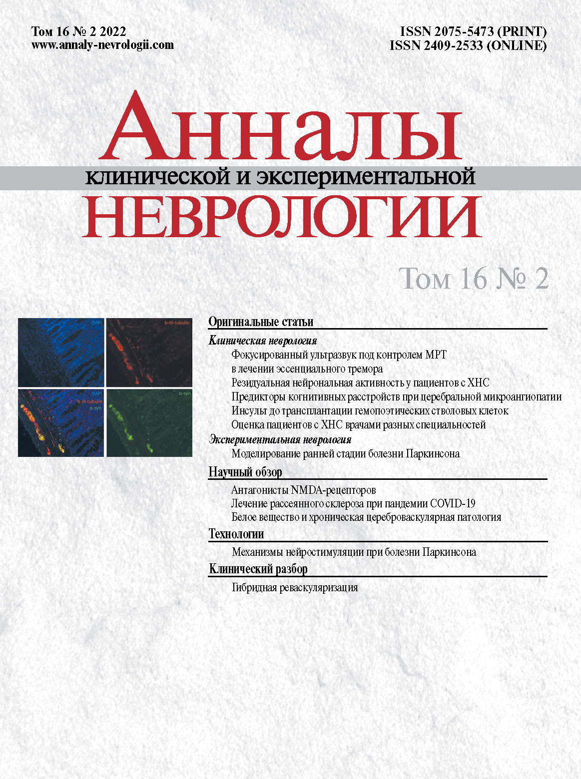
Vol 16, No 2 (2022)
- Year: 2022
- Published: 30.06.2022
- Articles: 11
- URL: https://annaly-nevrologii.com/journal/pathID/issue/view/73
Full Issue
Original articles
First use of MRI-guided focused ultrasound to treat patients with essential tremor in Russia
Abstract
Introduction. Treatment with MRI-guided focused ultrasound (MRgFUS) is a new, non-invasive surgical technique for treating extrapyramidal movement disorders. This article presents the first use of MRgFUS in Russia for treating patients with essential tremor (ET).
Materials and methods. Patients (n = 26; 17 men and 9 women) aged 21–82 years (median age 46.0 years) and with severe and refractory ET, underwent MRgFUS thalamotomy (ExAblate 4000, Insightec). One side was treated in 22 patients (left thalamus in 18 and right thalamus in 6), both sides were treated concurrently in two patients, and both sides were treated consecutively in two patients. Tremor was assessed using the Clinical Rating Scale for Tremor (CRST). Because international clinical specialists could not visit Russia due to the COVID-19 pandemic, MRgFUS was performed via telehealth on May 5, 2020, in a world first.
Results. A satisfactory result was achieved in 25 (96%) out of 26 patients. CRST scores improved by 64.7% on the side of the operation, by 10.2% on the control side, and by 37.5% overall. Intraoperative side effects included headache during sonication (42.3%), vertigo (15.4%), nausea (11.5%), vomiting (7.7%), numbness (3.8%), ataxia (3.8%), and pathological response to cold exposure (3.8%). The symptoms resolved immediately after surgery. Unstable gait was noted in five patients, which completely resolved two weeks after surgery. Median postoperative follow-up duration was 109 days [53; 231], with a maximum of 625 days. No relapses (if the hyperkinesia had completely disappeared) or increased tremor (if reduced after surgery) were observed.
Conclusion. The efficacy of MRgFUS for ET was 96%, with no long-term complications. Both bilateral concurrent and bilateral consecutive MRgFUS thalamotomy is possible, but its efficacy and safety should be assessed in a randomized study. In a world first, MRgFUS was successfully implemented using telehealth.
 5-14
5-14


Features of residual brain activity in patients with chronic disorders of consciousness on resting-state functional MRI
Abstract
Introduction. Rapid advances in critical care medicine have led to an increased survival rate of patients with severe brain damage and, consequently, to an increased prevalence of chronic disorders of consciousness (CDC). The lack of or fluctuations in signs of consciousness, which accompany the restoration of alertness after recovery from coma, indicate whether the type of CDC is a vegetative state or minimally conscious state. Correct diagnosis determines not only the rehabilitation outcome but also the economic outlook for a particular patient. However, the subjective nature of signs of consciousness, which are identified during clinical examination using neurological scales, is a common cause of diagnostic errors. The study of spontaneous activity using resting-state functional magnetic resonance imaging (fMRI) has helped to identify resting state networks. The default mode network (DMN) is one of the most studied brain networks. Its signal can change or be absent in patients with various types of CDC.
Purpose. To study the signal of residual spontaneous brain activity in patients with CDC at rest.
Materials and methods. Twenty-two patients with permanent CDC underwent resting state fMRI as an additional tool in the differential diagnosis between vegetative state and minimally conscious state at the Research Centre of Neurology.
Results. It was found that the nature of the signal coming from anatomical regions that are part of the DMN changes when signs of consciousness emerge.
Conclusion. These changes confirm that resting state fMRI is an important additional tool for differential diagnosis of CDC types. Accumulating knowledge about the brain's functional state helps us to expand our overall understanding of the nature of consciousness.
 15-24
15-24


Stroke before a haematopoietic stem cell transplantation is a potential risk factor for poor response to therapy in patients with blood cancer
Abstract
Introduction. More than 50,000 haematopoietic stem cell transplantations (HSCTs) are performed worldwide each year to treat malignant blood cancers, solid tumours, bone marrow aplasia, primary immunodeficiency conditions, autoimmune disorders, and storage disorders. The success of HSCTs depends on many factors, including patient's past medical history.
Purpose. To assess the effect of an acute cerebrovascular accident (CVA) that occurred before the HSCT on the transplantation outcome in patients with blood cancer.
Materials and methods. We examined the results of 899 transplantations conducted between 2016 and 2018 at the R.M. Gorbacheva Research Institute for Pediatric Oncology, Haematology and Transplantation of the Pavlov First Saint Petersburg State Medical University. We analysed transplantation parameters, as well as donor and recipient characteristics. Apart from intergroup comparisons, pseudo-randomization was performed using the Propensity Score Matching method. The survival rate analysis was conducted using the Kaplan–Meier estimate and the log rank test.
Results. Sixteen patients (1.8%) had cerebrovascular events in their past history before the HSCT: ischaemic stroke in 0.4% of cases and haemorrhagic stroke or intracerebral haemorrhage in 1.4% of cases. Patients with a history of cerebrovascular events included more people with leukaemia (p = 0.02), had more often received an allogenic transplant (р = 0.01), the donors more often had a partial rather than a full HLA match with the recipient (р = 0.06), had a lower body mass index (р = 0.02), and a lower Karnofsky/Lansky score (р = 0.01) than patients in the control group. The presence of a cardiovascular event had a statistically significant association with reduced overall survival rate of HSCT recipients (р = 0.0012).
Conclusion. Patients with blood cancer and stroke preceding the transplantation do not typically have any 'classical' risk factors (diabetes mellitus, venous system disorders, decreased cardiac output, significant atherosclerotic changes in precerebral arteries), therefore, secondary prevention guidelines for CVA during treatment of the main disease may not be effective and cannot be relied on. This article discusses the most likely causes of CVA in patients with blood cancer. A history of CVA before HSCT may have a significant effect on the transplantation outcome, but is not a contraindication for this treatment method. Recipient selection is a very important stage in HSCT planning. A multidisciplinary team should find a balance between the indications and contraindications for performing HSCT from an unrelated donor.
 36-43
36-43


Clinical assessment of patients with chronic disorders of consciousness by different medical specialists
Abstract
Introduction. Clinical assessment of consciousness in patients coming out of a coma remains a topic of discussion. Monitoring these patients over time is challenging not only because of the slow fluctuations in their neurological status, but also because doctors are not fully aware of the classification of chronic disorders of consciousness (CDC), and how to use the Coma Recovery Scale-revised (CRS-R), which was specifically developed for this group of patients. In practice, most doctors use standard neurological examination to assess consciousness rather than the CRS-R. We have hypothesized that this approach leads to contradictory and poorly standardized results.
Materials and methods. We investigated the level of inter-expert reliability in pairs of three medical specialists: neurologists, neurosurgeons and neurocritical care specialists (working in neurocritical care units) in the clinical assessment of consciousness. Their examination findings were compared to the CRS-R scores.
Results. The inter-expert reliability was poor in all three specializations when using clinical examination to determine the degree of impaired consciousness in patients with CDC. An average level of IER (Cohen's kappa = 0.46) was found only in the neurosurgeon–CRS-R pair.
Conclusion. A scale with detailed criteria is different to a standard clinical examination and has a higher level of IER. Moving from subjective evaluation to a standardized CRS-R will enable medical specialists to determine a patient’s rehabilitation potential and predict disease progression more accurately. Educational programmes, including virtual platforms, should be developed to encompass most of the medical community.
 44-49
44-49


Relations of impaired blood flow and cerebrospinal fluid flow with damage of strategic for cognitive impairment brain regiones in cerebral small vessel disease
Abstract
Introduction. Cerebral small vessel disease (CSVD), associated with age and vascular risk factors, as well as the main cause of vascular and degenerative mixed cognitive impairment (CI). Previously established microstructural predictors of CI (axial diffusion in normal-appearing periventricular white matter of the posterior left frontal lobe, the right midcingulate cortex, and the middle posterior part of the corpus callosum) can be used to calculate an integrative factor, exceeding the threshold value for which indicates the presence of CI. The use of this factor in the diagnosis of CI in CSVD is supported by the fact that leading mechanisms of CSVD are involved in the damage to areas of the brain that are strategic for CI.
The aim of this study was to clarify the link between the known microstructural predictors of CI in CSVD and MRI findings that correspond to the main mechanisms of CSVD.
Materials and methods. Patients (n = 74; including 48 women; average age 60.6 ± 6.9 years) with CSVD and CI of varying severity underwent phase-contrast MRI and voxel-based morphometry (3T) to assess arterial, venous and CSF flow, as well as atrophy.
Results. The established microstructural predictors of CI correlated with measures of arterial and venous blood flow, as well as atrophy. Linear regression models allow us to estimate cognitive impairment (CI) predictors in cerebral small vessel disease (CSVD), based on increased arterial velocity pulse index, CSF flow at the level of the cerebral aqueduct, cerebral aqueduct area and lateral ventricles volume, when there is reduced blood flow in the superior sagittal sinus and the overall arterial blood flow.
Conclusion. The ability to calculate microstructural predictors of CI due to CSVD, based on MRI findings, indicates the validity of using an integrative measure of microstructural predictors of CI as a diagnostic tool of CI in CSVD.
 25-35
25-35


Modelling motor and non-motor signs of early-stage Parkinson's disease
Abstract
Introduction. As Parkinson's disease (PD) develops, a number of non-motor signs precede motor symptoms, including gastrointestinal tract dysfunction. Modelling early-stage PD to comprehensively assess the pattern of morphofunctional changes in the gastrointestinal tract is important in order to develop methods of early disease diagnosis and more effective treatment of autonomic disturbances that are typical in PD, and to increase the patients' quality of life.
Study aim — to offer a model of early-stage PD through long-term oral administration of small doses of the neurotoxin rotenone to rats, and to study the functional and immunohistochemical changes in the gastrointestinal tract of the experimental animals, as well as changes in the substantia nigra.
Materials and methods. The experiment was conducted in male Wistar rats aged 3.0–3.5 months. The study group rats (n = 10) were given rotenone orally at a dose of 5 mg/kg, as a suspension in a 4% carboxymethyl cellulose solution, every second day for 7 months. The control group rats (n = 10) received only the 4% carboxymethyl cellulose solution.
The animals' mobility was assessed at the start and end of the experiment using the open field and narrowing beam-walking test. Gastrointestinal motility was assessed by measuring the passage of dye from the pylorus in a caudal direction along the small intestine. The rats were decapitated and immunohistochemistry was used to assess the density of dopamine neurons in the substantia nigra, nerve fibres, and glia in the Auerbach's plexus of the small intestine, and the location of the total and phosphorylated alpha-synuclein in the enteric nervous system.
Results. Rats in the study group had a statistically significant reduction in the number of dopamine neurons in the substantia nigra. Auerbach's plexus of the small intestine contained significantly less nerve fibres and glia, while fluorescence intensity for alpha-synuclein was increased. Phosphorylated alpha-synuclein was identified in the cholinergic and adrenergic fibres of Auerbach's plexus. Experimental animals had a statistically significant reduction in the gastric emptying rate and small intestine motility compared to the control group.
Conclusion. The presented model of early-stage PD enables the physiological and immunohistochemical symptoms of gastrointestinal dysfunction, similar to that of patients with PD, to be replicated. They are based on intestinal denervation changes and accumulation of abnormal forms of alpha-synuclein in the enteric nervous system.
 50-57
50-57


Reviews
NMDA receptor antagonists as potential therapy in cerebellar degenerative disorders
Abstract
Cerebellar degeneration remains a poorly studied topic. Excitotoxicity, i.e. neuronal damage and death due to excess activation of postsynaptic N-methyl-D-aspartate receptors (NMDAR) by glutamate, is considered to be a universal mechanism of most neurodegenerative conditions. The use of antagonists that predominantly block NMDAR in cases of excitotoxicity is a very promising treatment strategy for neurodegenerative disorders.
This review presents the known structure and function of NMDAR. Information on studies investigating the use of NMDAR antagonists in the treatment of neurodegenerative diseases is provided. Creation of new therapies to correct excitotoxicity in various neurodegenerative disorders, for example, spinocerebellar ataxias, requires further study of the subunit composition and the role of NMDAR in the cerebellum. Treatment methods that combine the use of extrasynaptic NMDAR antagonists or synaptic NMDAR agonists with drugs that affect the total amount of glutamate in the synaptic cleft are promising.
 58-69
58-69


Safety of pathogenetic therapy for multiple sclerosis during the COVID-19 pandemic
Abstract
The safety of pathogenetic therapy for multiple sclerosis (MS) is a crucial aspect of the therapeutic strategy during the COVID-19 pandemic.
Based on our own data, obtained during the study of MS pathogenesis and safety analysis of MS disease-modifying therapies (DMTs), we hereby suggest a classification of DMTs side effects, based on their type, development, and direction of action. There is a need to thoroughly analyse adverse events caused by pathogenetic therapy, with a balanced assessment of the direct vs. adverse effects of immunosuppressive drugs.
Based on available literature, in the article, data on the effect of DMTs with various mechanisms of action on severe coronavirus infection are systematized.
Interferon-β and glatiramer acetate are the safest drugs to use during the COVID-19 pandemic. Teriflunomide, dimethyl fumarate, natalizumab, ocrelizumab, fingolimod, alemtuzumab, and cladribine should be used with caution. Drugs with a minor systemic immunosuppressant effect (e.g. natalizumab) and selective immunosuppressants (e.g. ocrelizumab) are safer than drugs that cause non-selective depletion of T and B cells.
It must be stressed that the risk of MS exacerbation and progression due to untimely prescription or cessation of pathogenetic therapy can significantly exceed the potential risk of COVID-19.
Long-term safety monitoring is required for DMTs during the COVID-19 pandemic and when the epidemiological situation stabilizes.
 70-77
70-77


Morphology and pathogenesis of white matter changes in chronic cerebrovascular disease
Abstract
This review contains up-to-date information on the fundamentals and clinical aspects of white matter disease in chronic progressive cerebrovascular disease with cognitive impairment, the leading risk factors for which are hypertension and cerebral atherosclerosis. Highly informative methods of neuroimaging have contributed significantly to the study of this problem, confirming the important role of white matter changes in the development and progression of cognitive impairment. The full range of the morphological changes in white matter, typical of vascular encephalopathy and cognitive dysfunction, is presented. Chronic hypoperfusion and white matter ischaemia play a leading role in the pathogenesis of white matter changes in vascular dementia development, but alternative hypotheses are also emerging. Further fundamental morphological and clinical studies will help to determine the leading mechanisms of white matter damage in patients with vascular and other age-related forms of dementia. This is necessary for the development of effective methods of treatment and prevention.
 78-88
78-88


Technologies
Current understanding of neurostimulation for Parkinson's disease
Abstract
Specific mechanisms underlying the therapeutic effects of neurostimulation in Parkinson's disease remain a topic of discussion and intense study. Understanding these mechanisms can serve as the foundation for developing and selecting more effective parameters to relieve the symptoms of Parkinson's disease, maximize the advantages, and reduce the adverse effects and need for surgical intervention. The article discusses existing models of motor control in the basal ganglia in healthy individuals and in PD from the point of view of neuromodulation (changes in the impulse flow model, oscillatory model), as well as the current understanding of the mechanisms of action of deep brain stimulation (DBS): the block depolarization hypothesis, neural interference hypothesis, synaptic depression hypothesis, synaptic modulation hypothesis, and the DBS astrocytes hypothesis. Factors such as DBS location and neurostimulation parameters, affecting the clinical outcome, are considered in detail. The neuroprotective effect of DBS is also touched on.
 89-99
89-99


Clinical analysis
Hybrid revascularization: stenting of left internal carotid artery after restenosis, combined with left anterior descending artery stenting and right carotid endarterectomy
Abstract
We present a description of internal carotid artery restenosis five years after eversion carotid endarterectomy, progression of contralateral internal carotid artery stenosis and coronary atherosclerosis with imaging of haemodynamically significant stenosis of the anterior descending artery. The efficacy of hybrid revascularization is demonstrated: carotid angioplasty with stenting of the left internal carotid artery stenosis + percutaneous coronary intervention for the anterior descending artery + left carotid endarterectomy. We explain the choice of these types of reconstruction and staging of the intervention. We make a conclusion regarding the effectiveness and safety of the implemented treatment strategy.
 100-105


