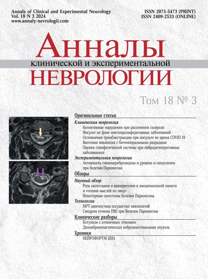
Vol 18, No 3 (2024)
- Year: 2024
- Published: 03.10.2024
- Articles: 13
- URL: https://annaly-nevrologii.com/journal/pathID/issue/view/83
Original articles
Spectrum of Cognitive Impairment in Patients with Multiple Sclerosis
Abstract
Introduction. Cognitive impairment (CI) is a common manifestation of multiple sclerosis (MS), which significantly affects patients’ daily life and professional activity. Despite the development of methods to screen MS patients for CI, data on its prevalence in the Russian population are still lacking.
Aim: to comprehensively assess cognitive functions in patients with different types of MS.
Materials and methods. The study included MS patients who did not have any other possible causes of CI and no diseases or conditions that confounded this assessment. CI was determined using the Brief International Cognitive Assessment in Multiple Sclerosis (BICAMS) test battery and the Stroop test as a decrease in the scores below the mean by at least 1.5 standard deviations. CI was subjectively assessed using the Perceived Deficit Questionnaire; fatigue was subjectively assessed using the Modified Fatigue Impact Scale (MFIS). The Mann–Whitney test and Fisher’s exact test were used for comparison, and the Spearman test was used to evaluate correlations.
Results. We evaluated 77 MS patients (30 men; age 40 [30; 48] years; 47 with relapsing-remitting MS, 30 with progressive MS). CI incidence was 23.4% in patients with relapsing-remitting MS and 77% in patients with progressive MS, while multi-domain CI was statistically significantly more common in patients with progressive MS. Impairment of processing speed was the most common. Patients with relapsing-remitting MS and CI were statistically significantly older and had longer disease duration than those without CI. There was a statistically significant correlation of subjective CI severity with MFIS scores but not with testing results.
Conclusion. CI incidence in MS patients was relatively high with greater severity and involvement of more domains in patients with progressive MS. No correlation was found between subjective and objective CI assessment results, which may suggest that patients underestimated their deficit.
 5-13
5-13


Clinical and Neuroimaging Patterns of Ischemic Stroke in Ph-negative Myeloproliferative Neoplasms
Abstract
Introduction. Philadelphia-negative myeloproliferative neoplasms (MPNs) are a rare blood disorder characterized by pancytosis and thrombohemorrhagic complications.
The aim of this article is to describe clinical and neuroimaging patterns of brain changes in patients with MPN.
Materials and methods. The study included 152 patients with an established diagnosis of MPN (according to WHO criteria 2008, 2016). A clinical and neurological examination, laboratory tests, and magnetic resonance imaging of the brain were performed.
Results. In patients with polycythemia vera and primary myelofibrosis, neuroimaging patterns are represented by small (up to 1.5 cm) post-infarction lesions in the brainstem, cerebellum, and cortex in adjacent perfusion territories after hemorheological microocclusive stroke. In patients with essential thrombocythemia, the neuroimaging pattern is more often represented by massive post-infarction changes in cortical-subcortical brain tissue with atherosclerotic lesions of the major head arteries, which appear to be atherothrombotic. Stroke preceded hematologic diagnosis in 30% of polycythemia vera cases, 40% of essential thrombocythemia cases, and 25% of primary myelofibrosis cases.
 14-25
14-25


Ischemic Stroke and Coronavirus Infection: Complications of Endovascular Thrombectomy
Abstract
The objective of our study was to compare complications of endovascular thrombectomy (EVT), in ischemic stroke (IS) patients admitted with or without COVID-19 to hospitals converted to deliver COVID-19-specific care.
Materials and methods. A retrospective analysis of 817 clinical cases of IS patients aged 25–99 years treated in regional vascular centers of Saint Petersburg from 1 January to 31 December 2021, with confirmed thrombotic occlusion of cerebral vessels and subsequent EVT intervention.
Results. The EVT number per bed was significantly higher (1.6) in the non-converted hospitals compared to the COVID-19-converted hospitals (0.49; p < 0.001). At the same time, more intraoperative complications (12% vs. 7.1%; p = 0.03) were reported in non-converted hospitals compared to COVID-19 converted hospitals. The likelihood of a favorable functional outcome was higher in younger patients with less severe neurological deficits on admission and without concomitant COVID-19 or post-operative complications.
Conclusion. COVID-19 is a limiting factor for the effectiveness of an IS treatment in patients who underwent EVT, affecting thereby functional outcomes in this cohort of patients. The impact of the COVID-19 pandemic on intra-operative EVT complication rate was associated with disrupted triage of IS patients and an uneven distribution of the workload among surgical teams in the city hospitals.
 26-34
26-34


Temporal Lobe Epilepsy with Bitemporal Interictal Epileptiform Discharges: Effects of Sleep and Wakefulness
Abstract
Introduction. Independent bitemporal interictal discharges are often found in patients with temporal lobe epilepsy. The likelihood of registering epileptiform activity (EA) is higher during sleep. Assessment of bitemporal interictal epileptiform discharges (BIEDs) with various discharge predominance ratio is used for presurgical evaluation of epilepsy patients and prediction of surgical outcomes.
Our objective was to determine the predominant side (PS) in patients with bitemporal epilepsy using the incidence of epileptiform discharges for each sleep stage.
Materials and methods. We analyzed 45 recordings of 10–24 h long-term video-EEG monitoring (LTM) in patients with bitemporal EA. For each recording, the total incidence of EA (IEA) and EA incidence for wakefulness and for each sleep stage were calculated individually. We also assessed the discharge predominance index (DPI) as a ratio of IEA in the predominant and contralateral sides for the entire recording and for each sleep stage.
Results. We observed an IEA increase with sleep deepening, with maximum values observed during N2 and N3 sleep stages. The minimum IEA values were recorded during REM sleep; nevertheless, most of the REM sleep discharges were detected on the PS. DPI values were the highest and the most stable during N2 and N3 stages.
Conclusion. The findings of our study demonstrate an increase in DPI values with non-rapid eye movement (NREM) sleep deepening in patients with bitemporal localization of EA. Despite the protective effects of REM sleep (i.e., reducing the likelihood of EA), it may be pivotal in lateralization of EA in patients with BIEDs. The PS is generally determined by a higher DPI during N2 and N3 stages.
 35-41
35-41


Glymphatic System Assessment Using DTI-ALPS in Age-Dependent Neurodegenerative Diseases
Abstract
Introduction. Dysfunction of the glymphatic system glymphatic system of the brain is considered a pathogenetic factor in some age-dependent neurodegenerative diseases, including Alzheimer's disease (AD), dementia with Lewy bodies (DLB), Parkinson's disease (PD), and normal pressure hydrocephalus (NPH). The innovative method for calculating DTI-ALPS (Diffusion Tensor Image Analysis ALong the Perivascular Space) allows non-invasive assessment of the glymphatic system status using magnetic resonance imaging (MRI).
The aim of the study is to compare DTI-ALPS in patients with AD, DLB, PD, and NPH and to evaluate its potential use as a biomarker of the glymphatic system status in these diseases.
Materials and methods. The study included 116 subjects: 32 patients with AD, 15 patients with DLB, 31 patients with PD, 11 patients with NPH, and 27 healthy volunteers. Cognitive testing was performed for patients in the main groups using the Montreal Cognitive Assessment (MoCA) score. All subjects underwent diffusion tensor imaging (DTI) of the brain. DTI-ALPS was then calculated.
Results. DTI-ALPS index significantly differed across groups (p < 0.001). Patients with AD, DLB, and NPH had a significantly lower DTI-ALPS index on both sides compared to the PD group and healthy volunteers (p < 0.01). Analysis of the entire sample showed a direct correlation between MoCA score and DTI-ALPS index (p < 0.05).
Conclusion. This is the first comparison of DTI-ALPS across such a broad range of age-dependent neurodegenerative diseases. Since our DTI-ALPS results were comparable to previously reported data, we believe that this parameter can be used as an indirect marker of the glymphatic system status.
 42-49
42-49


Blood Glucocerebrosidase Activity and α-Synuclein Levels in Patients with GBA1-Associated Parkinson's Disease and Asymptomatic GBA1 Mutation Carriers
Abstract
Introduction. Mutations in a GBA1 gene, which encodes a lysosomal enzyme called glucocerebrosidase (GCase), are the most common genetic risk factor for Parkinson's disease (PD). The pathogenesis of PD results from the death of dopaminergic neurons in the substantia nigra of the brain, which is associated with the aggregation of α-synuclein protein. However, not all GBA1 mutation carriers develop PD during their lifetime.
The aim of this study was to evaluate GCase activity and α-synuclein levels in CD45+ blood cells of patients with PD associated with GBA1 mutations (GBА1-PD), asymptomatic carriers of GBA1 mutations (GBА1-carriers), and patients with sporadic PD (sPD), as well as correlation between the study parameters in the study groups.
Materials and methods. The study included patients with GBА1-PD (n = 25) and sPD (n = 147), and GBА1-carriers (n = 16). A control group included healthy volunteers (n = 154). The level of α-synuclein in CD45+ cells was measured by enzyme-linked immunosorbent assay, and GCase activity in dried blood spots was detected by high-performance liquid chromatography with tandem mass spectrometry.
Results. Increased level of α-synuclein protein was detected in CD45+ blood cells of patients with GBA1-PD, sPD, and GBA1-carriers compared to controls (p = 0.0043; p = 0.0002; p = 0.032, respectively). Decreased GCase activity was reported in GBA1-PD patients and GBA1-carriers compared to sPD patients (p = 0.0003; p = 0.003, respectively) and controls (p < 0.0001; p < 0.0001, respectively). However, negative correlation between α-synuclein levels and GCase activity was observed only in GBA1-PD patients, but not in GBA1-carriers.
Conclusion. Our data suggest a possible functional relationship between the activity of GCase and the metabolism of α-synuclein in PD associated with GBA1 mutations.
 50-57
50-57


Reviews
Oxytocin and Vasopressin in Emotional Memory and “Face Reading”: a Neurobiological Approach and Clinical Aspects
Abstract
The ability to adequately perceive and recognize emotions is a key and universal tool in interpersonal communication, which allows people to understand feelings, intentions, and emotional reactions of themselves and others. Throughout their life, people have to make inferences about mental state of others by interpreting subtle social signals, such as facial expressions, to understand or predict others’ behavior, which is crucial in constructive social interactions. Therefore, emotional memory associated with the ability to identify emotions based on one’s life experience is the cornerstone of social cognition and interpersonal relationships. Oxytocin and vasopressin are neurohypophysial peptides that have attracted scientific attention due to their role in the emotional and social aspects of behavior. Variable and contrasting effects of oxytocin and vasopressin may be related to the sites of the brain where they exert their activity.
Aim. This review aimed to evaluate neural mechanisms underlying oxytocin-mediated and vasopressin-mediated modulation of emotional memory; to assess how cerebral oxytocin-signal and vasopressin-signal transduction mediates emotional and social behavior; to discuss the role of the two neuropeptides in non-verbal interpersonal communication; and to present their cerebral effects in relation to an ability for “face reading” in patients with mental disorders.
 58-71
58-71


Spectrum of Non-Motor Symptoms in Parkinson’s Disease — a Review
Abstract
Motor and non-motor symptoms of Parkinson’s disease (PD) and their management have been evaluated in numerous studies. Four classical symptoms, including bradykinesia, tremor, rigidity, and postural abnormalities, are used to establish a clinical diagnosis of PD. However, this research is aimed at exploring the range of non-motor symptoms with an emphasis upon their ability to affect the patients with PD and their quality of life.
With a slow onset of the known symptoms like tremor or rhythmic shaking of limbs called “pill-rolling tremor”, slowed movement (bradykinesia), muscle rigidity, stooped and altered posture, loss of the ability to blink or smile, and various speech and writing changes; the disease takes a leap into the non-motor symptoms like dementia, drooling, swallowing issues, difficulty urinating, and constipation. The dopaminergic pathophysiology of PD explains the anxiety, slowness of thought, fatigue, and dysphoria. Knowing the non-motor symptoms is crucial to help the clinician to make early diagnosis and to better understand the prognosis of the spectrum of this disease.
 72-80
72-80


Magnetic Resonance Imaging Diagnostics of Vascular Myelopathies: from Basic Sequences to Promising Imaging Protocols
Abstract
Magnetic resonance imaging (MRI) is the method of choice in diagnostics and differential diagnosis of spinal cord arterial infarction and venous insufficiency. However, imaging of vascular myelopathy is complicated by the lack of clear diagnostic criteria. Basic MRI sequences have low sensitivity at disease onset, and described MR patterns do not sufficiently increase imaging specificity for spinal cord ischemia, so imaging protocols are to be elaborated.
Diffusion-weighted imaging is a key additional sequence that allows establishing the ischemic nature of myelopathy.
Inclusion of spinal MR angiography in comprehensive MR examination allows visualization of aorta abnormalities, its large branches or spinal arteriovenous fistulas, so that they can be treated early.
We presented an optimal MRI protocol for patients with suspected ischemic spinal stroke. Promising high-tech MR sequences for visualization of vascular myelopathies were reviewed.
 81-90
81-90


Deep Brain Stimulation Withdrawal Syndrome, a Rare Life-Threatening Condition in Neurology and Neurosurgery
Abstract
The article addresses an acute condition associated with an abrupt cessation of neurostimulation of deep brain structures, which is manifested by acute hypokinesia and rigidity with further development of akinesia, anarthria and dysphagia. This may result in the need for emergency hospitalization and admission to an intensive care unit. The article presents literature review and clinical case reports. We discuss causes and approaches to the prevention and management of acute decompensation in patients with Parkinson's disease associated with abrupt deep brain stimulation cessation.
 91-102


Clinical analysis
Clinical Case of Atypical Botulism with Pseudointernuclear Ophthalmoplegia Syndrome
Abstract
Botulism is a rare cause of bulbar and oculomotor syndromes. A late diagnosis and, therefore, late initiation of specific therapy may lead to multiple life-threatening complications. Epidemiological history and clinical findings are key to the correct diagnosis, but if these data are not available due to atypical clinical findings, botulism identification is challenging.
In our clinical case, a 31-year-old man was admitted to the hospital with double vision, impaired eye movements, and difficulty swallowing rapidly developing for 2 days. Ocular motility dysfunction included disturbed conjugate eye movements. In young patients, this is most often caused by demyelinating disease with medial (posterior) longitudinal fasciculus damage and symmetrical bilateral ptosis. The patient denied eating foods that could cause botulism and did not have any gastrointestinal symptoms. Differential diagnoses included demyelinating disease onset and Miller–Fisher syndrome. The next morning, completely identical clinical signs appeared in the patient’s mother who had eaten canned mushrooms, so botulism was suspected. Over the next few hours, despite the administration of anti-botulinum serum, acute respiratory failure developed, and the patient was placed on a ventilator for 28 days. The patient and his mother were discharged in a satisfactory condition, and their symptoms completely resolved within a few months. The diagnosis of botulism was confirmed by toxicological examination.
 103-108


Atypical Presentation of Dysembryoplastic Neuroepithelial Tumor
Abstract
Dysembryoplastic neuroepithelial tumor (DNET) is a benign glioneuronal neoplasm, usually found in children and adolescents, in the vast majority of cases associated with drug-resistant epilepsy. Typically, epileptic seizures are the main, and in most cases, their only clinical manifestation. Although DNET is a benign, biologically stable tumor with few reports of malignancy, it is one of the most common reasons for epileptic surgery. The epileptogenic potential of this tumor is so high that DNET s, along with ganglioglioma, have received the informal term “epileptomas” and are by far the leaders in the group of low-grade tumors associated with long-term epilepsy-associated tumors (LEAT). It is believed that this epileptogenicity is due to localization in the neocortex and frequent association with focal cortical dysplasias (FCD). In the world literature, there are only a few mentions of DNET s not associated with epilepsy. The article presents the experience of complex, interdisciplinary diagnosis of DNET in a child without epilepsy who complained of frequent headaches. During a comprehensive MRI examination, a cortical-subcortical pathological substrate was discovered in the left temporal lobe with radiological signs of DNET. During video-EEG monitoring of night sleep, no epileptiform signs were recorded. There was no history of epileptic seizures or other paroxysms. A control MRI revealed a slight increase in the size of the pathological substrate, which was the reason for surgical treatment. Pathological examination revealed microscopic features of DNET. This case of absence of epilepsy in a child with cortical DNET in the temporal lobe cortex suggests that the spectrum of its clinical manifestations and biological behavior is not fully understood and requires further comprehensive study.
 109-115


Chronicle
NEUROFORUM-2024 is a key event in the field of neurology and neurosciences
 116-117


